Latin: nervus vestibularis
Overview
- The balance part of the vestibulocochlear nerve.
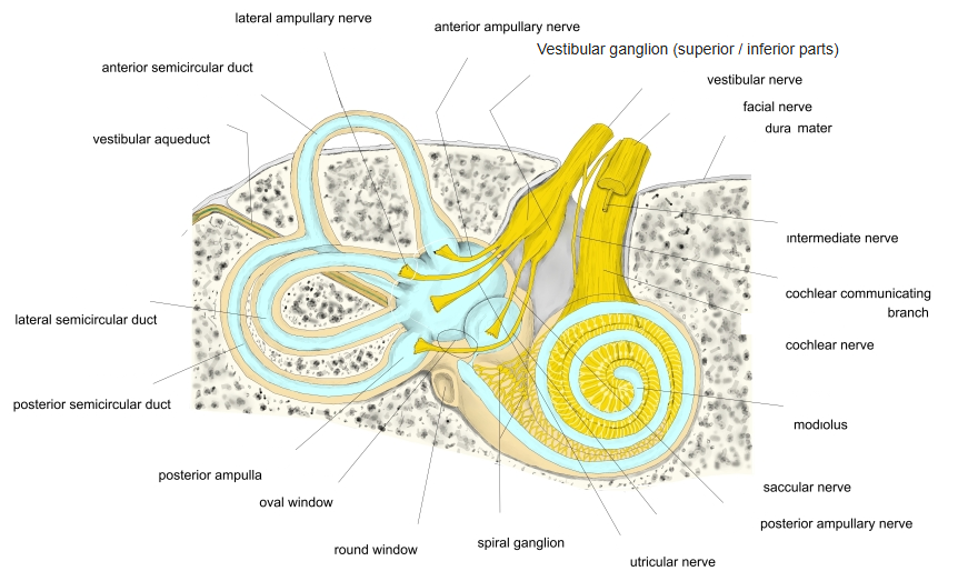
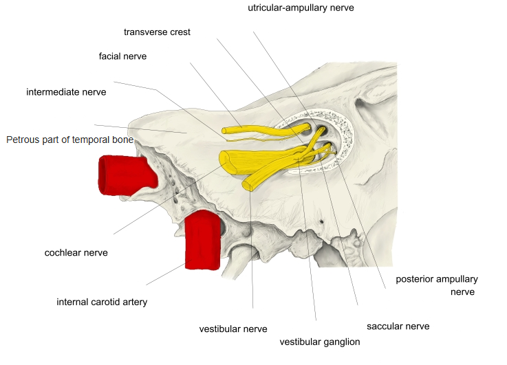
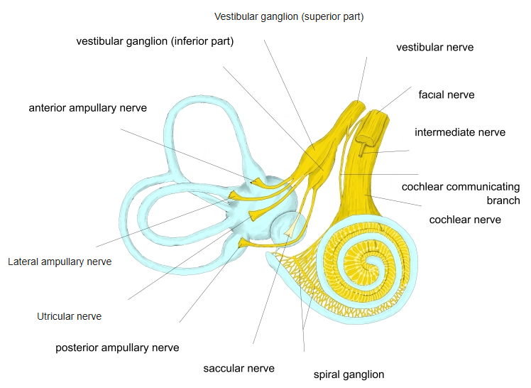
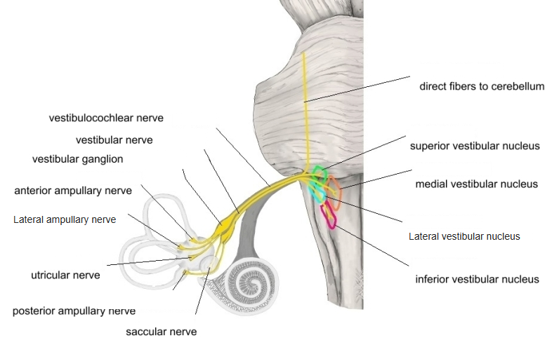
- Its sensory neurons form the vestibular ganglion in the fundus of the internal acoustic meatus; their central fibres enter the brainstem between the pons and the olive and end in the four vestibular nuclei (medial, lateral, superior, inferior).
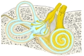
Inner ear and vestibulocochlear nerve 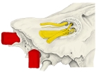
Internal acoustic meatus
Branches
| Part | Branches |
|---|---|
| Upper part (utriculo-ampullary nerve) | Utricular, anterior ampullary, lateral ampullary nerves |
| Lower part | Saccular nerve, posterior ampullary nerve |
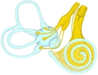
Branches of the vestibular and cochlear nerves 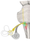
Vestibular nerve and vestibular nuclei 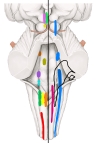
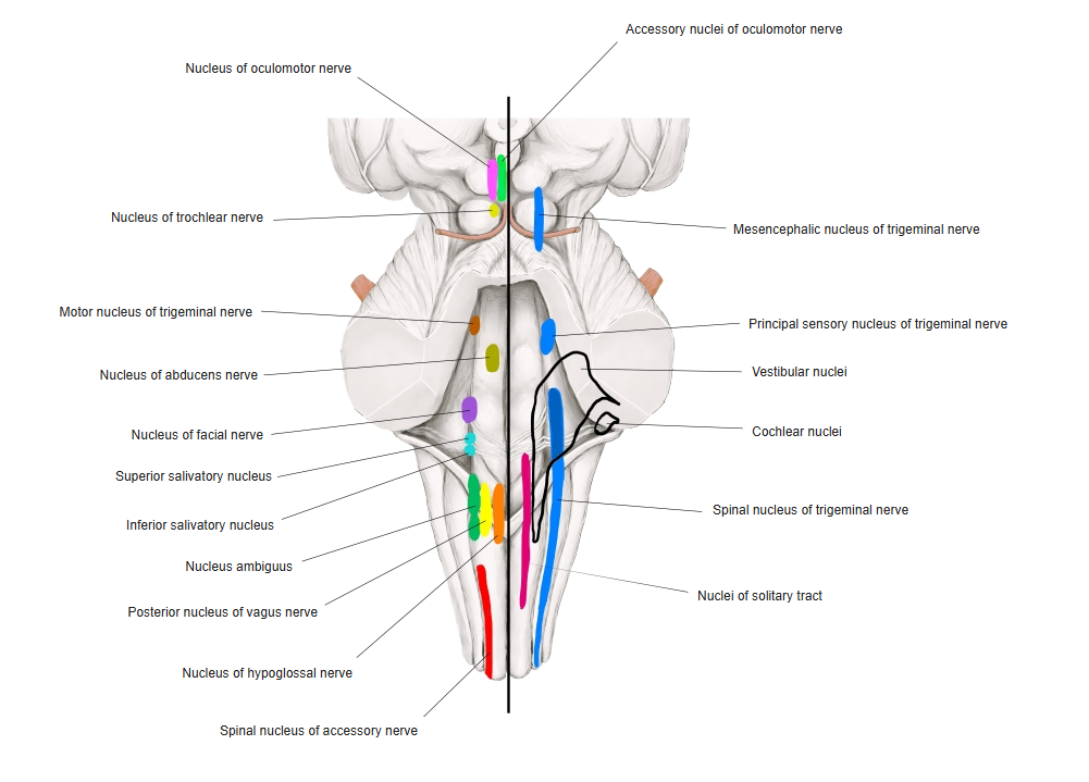
Cranial nerve nuclei in the brainstem
References and sources
- Further reading: Vestibular nerve, Wikipedia.