Latin: musculus abductor digiti minimi manus
Overview
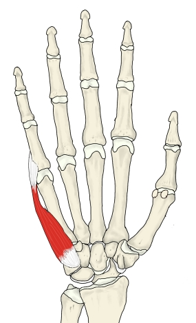
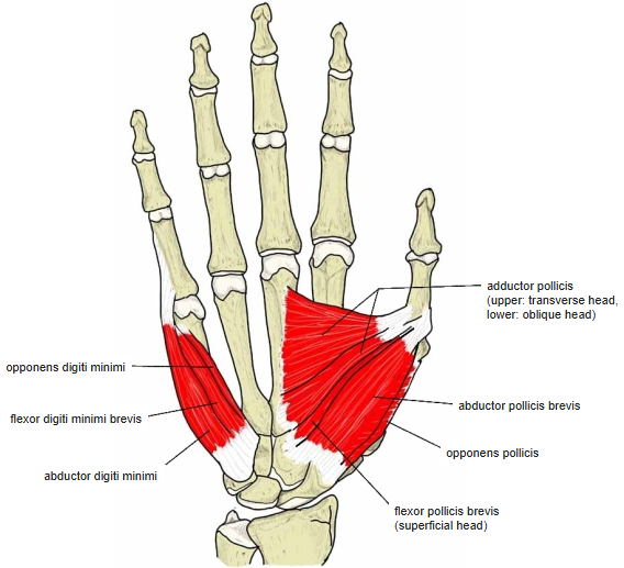
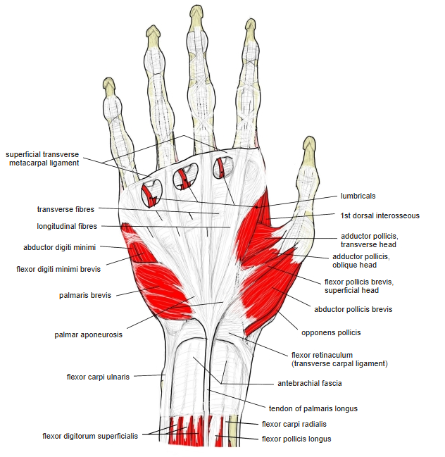
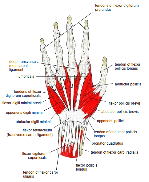
- One of the 4 hypothenar muscles (with the palmaris brevis, flexor digiti minimi brevis and opponens digiti minimi).
- Lies along the ulnar border of the palm; its spindle-shaped belly, with the skin and fat over it, forms the hypothenar eminence.
- It is the most superficial muscle of the hypothenar eminence. When the thumb and little finger are pressed together the hypothenar bellies are easy to feel, but distinguishing it takes practice, because abducting the little finger tightens the whole eminence.
- Develops early from the superficial layer of the ulnar muscle primordium of the hand (together with the flexor digitorum superficialis and abductor pollicis brevis); the other hypothenar muscles form later from the deep layer.
Position in the classification
Upper Limb › Hand › Hypothenar muscles
- Palmaris brevis
- Abductor digiti minimi (hand)
- Flexor digiti minimi brevis (hand)
- Opponens digiti minimi (hand)
Illustrations
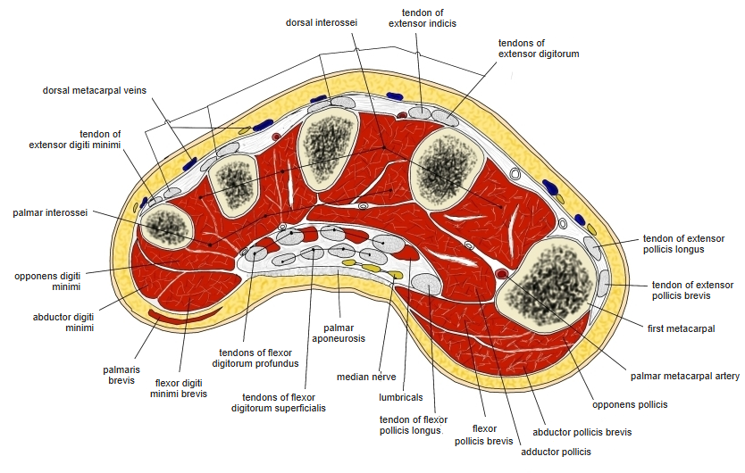
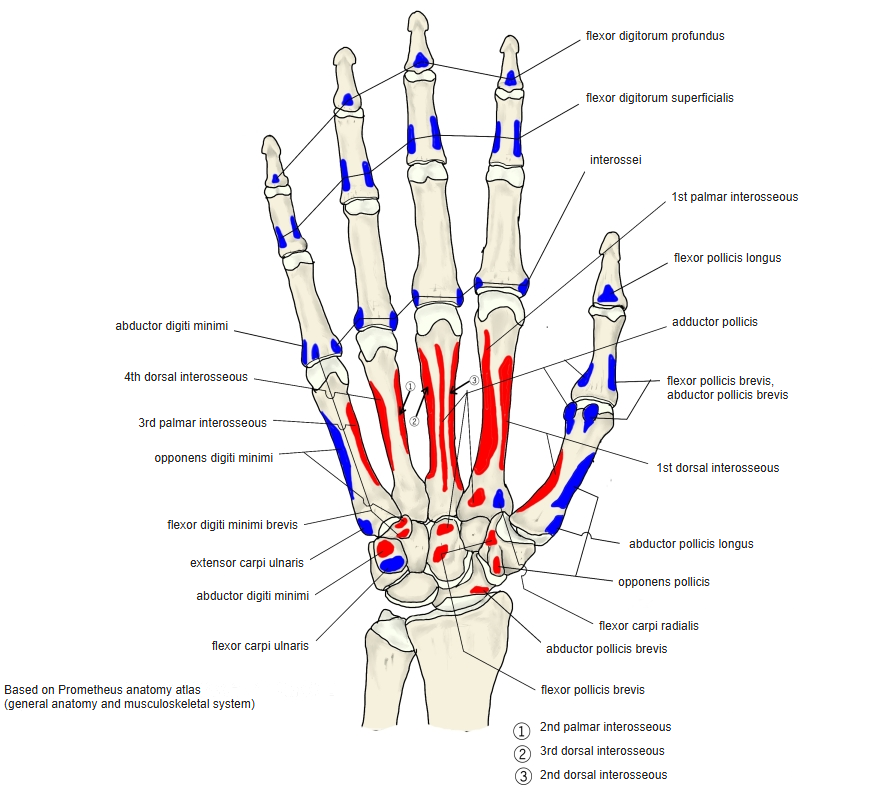
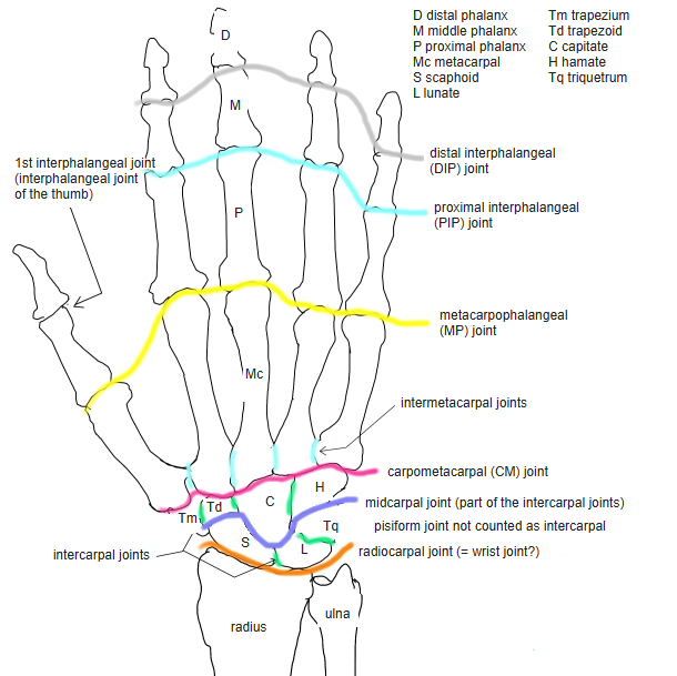
Origin / insertion
- Origin (sources differ):
- Pisiform (Prometheus atlas)
- Pisiform, pisohamate ligament and flexor retinaculum (other sources)
- Insertion (sources differ):
- The tendon divides into three slips: to the sesamoid bone on the ulnar side of the base of the proximal phalanx of the little finger (when present), to the palmar plate of the metacarpophalangeal joint, and to the margin of the anterior surface of the 5th metacarpal
- Ulnar side of the base of the proximal phalanx of the little finger and the dorsal digital expansion (Prometheus atlas and others)
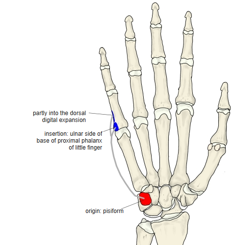
Action
- Abducts the little finger at the metacarpophalangeal joint
- Prometheus atlas: at the metacarpophalangeal joint flexes and abducts the little finger; at the interphalangeal joints extends it (through the dorsal digital expansion)
- Important when grasping large objects with the fingers spread
Innervation
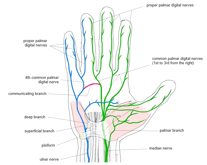
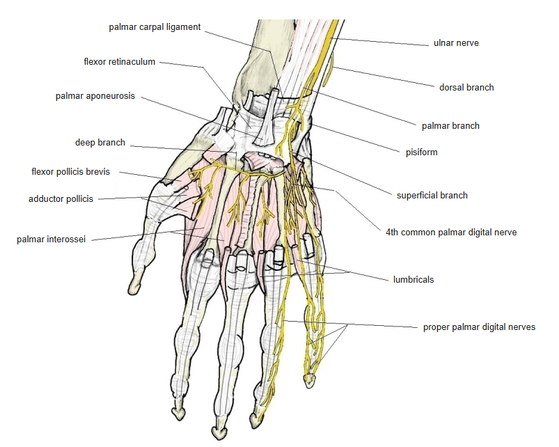
- Deep branch of the ulnar nerve (C8–T1)
Blood supply
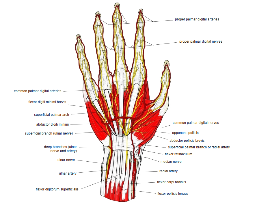
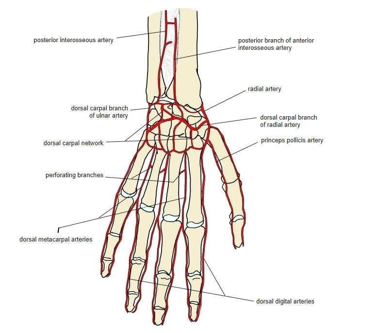
- Deep palmar branch of the ulnar artery
Anatomical variations
- The most variable hypothenar muscle
- Accessory slips may come from the antebrachial fascia, the radius, the ulna, the tendon of the flexor carpi ulnaris, the flexor retinaculum or the tendon of the palmaris longus
- Occasionally inserts partly on the 5th metacarpal
- With an extra (sixth) finger it may insert on that finger instead
Related terms
- Hypothenar muscles
- Flexor digiti minimi brevis (hand)
- Opponens digiti minimi (hand)
- Palmaris brevis
- Abductor digiti minimi (foot)
- Pisiform
- Pisohamate ligament
- Flexor retinaculum
- Dorsal digital expansion
- Ulnar nerve
- Ulnar artery
References and sources
- Further reading: Abductor digiti minimi muscle of hand, Wikipedia.