Latin: Venae ciliares anteriores
Overview
- Small veins at the front of the eyeball that accompany the anterior ciliary arteries.
- Receive blood from the front of the ciliary body and aqueous humour from the scleral venous sinus (canal of Schlemm), through the aqueous veins.
- Run back with the tendons of the rectus muscles and end in the muscular veins and the superior ophthalmic vein.
Veins at the corneoscleral junction
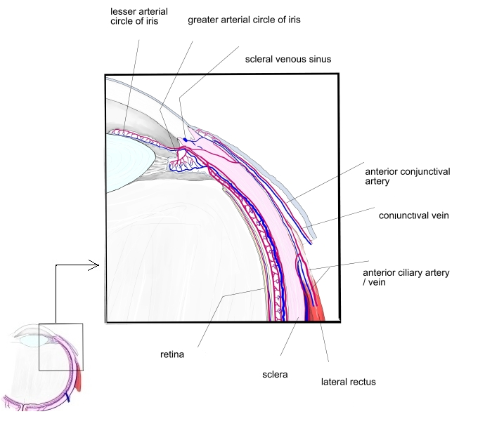
Tributaries and drainage
- Also receive the episcleral and conjunctival veins.
Clinical notes
- Their congestion around the cornea (ciliary flush) is a sign of inflammation inside the eye.
References and sources
- Further reading: Anterior ciliary veins, Wikipedia.