Latin: Plexus venosus vertebralis internus anterior
Overview
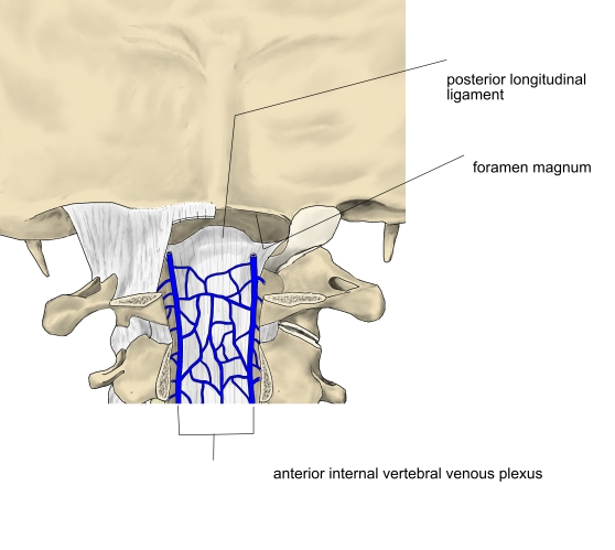
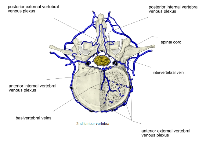
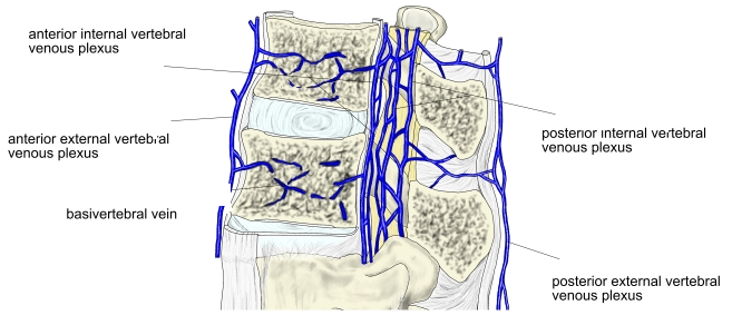
- Part of the internal vertebral venous plexus, in the epidural space of the vertebral canal.
- Consists of longitudinal veins on the backs of the vertebral bodies and discs, on each side of the posterior longitudinal ligament, linked by cross channels.
- Receives the basivertebral veins and drains through the intervertebral veins.
Anterior internal vertebral venous plexus 
Vertebral venous plexuses (cross section)
Connections
- Continuous above with the basilar plexus and the occipital sinus through the foramen magnum.
Vertebral venous plexuses (sagittal section) Vertebral venous plexuses (cross section)
Clinical notes
- The vertebral venous plexuses have no valves and connect the pelvic, abdominal and thoracic veins with the dural sinuses (Batson's plexus), a route for the spread of cancer (for example from the prostate or breast) and of infection to the vertebrae and brain.
References and sources
- Further reading: Anterior internal vertebral venous plexus, Wikipedia.