Latin: Venae vorticosae
Overview
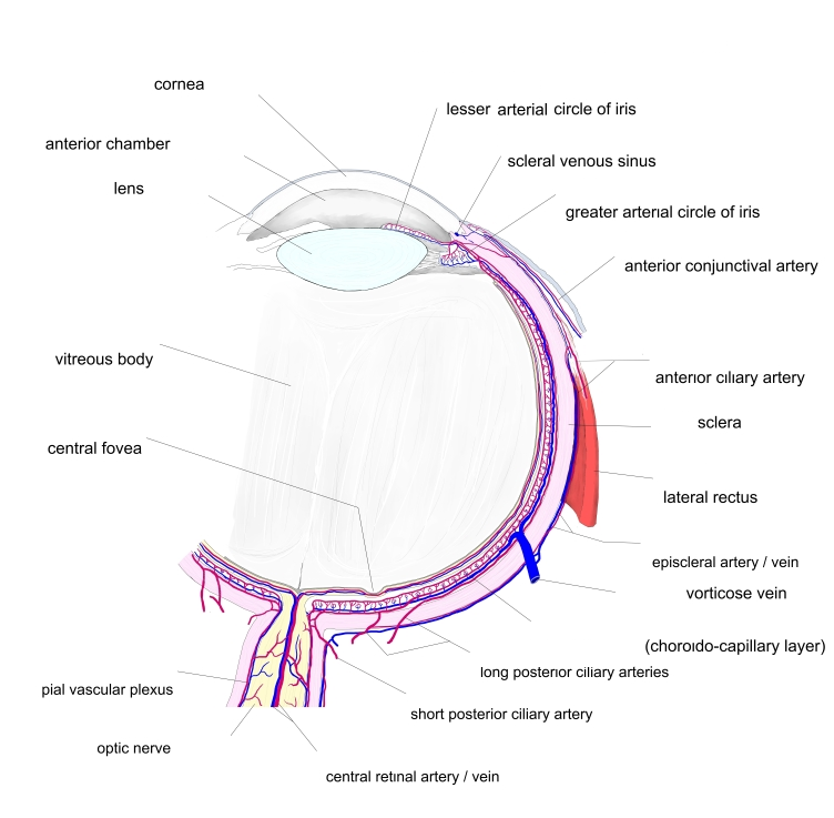
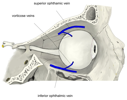
- The main veins of the choroid of the eyeball. Usually four (one in each quadrant), sometimes more.
- Formed by veins converging in a whorl in the choroid, then pass obliquely through the sclera behind the equator of the eye.
- The two upper veins end in the superior ophthalmic vein and the two lower in the inferior ophthalmic vein.
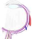
Blood vessels of the eyeball 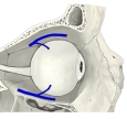
Vorticose veins
Tributaries and drainage
- Drain the choroid, most of the ciliary body and the iris.
References and sources
- Further reading: Vorticose veins, Wikipedia.