Latin: Venae hepaticae
Overview
- Carry all the blood that has passed through the liver into the inferior vena cava.
- Three main veins — right, middle and left — run between the segments of the liver (intersegmental), unlike the portal vein and hepatic artery, which run inside them.
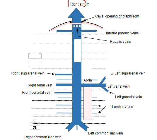
- Open into the IVC just below the diaphragm; the middle and left veins usually join first.
- Have no valves; several small veins from the caudate lobe enter the IVC directly.
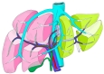
Hepatic veins and segments of the liver
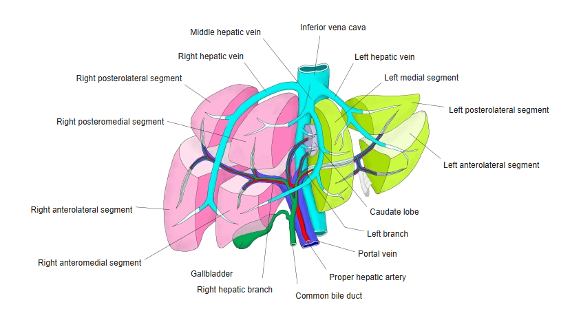
Main hepatic veins
| Vein | Position |
|---|---|
| Right hepatic vein | Between the right anterior and right posterior sections |
| Middle hepatic vein | Between the right and left halves of the liver (Cantlie line) |
| Left hepatic vein | Between the left medial and left lateral sections |
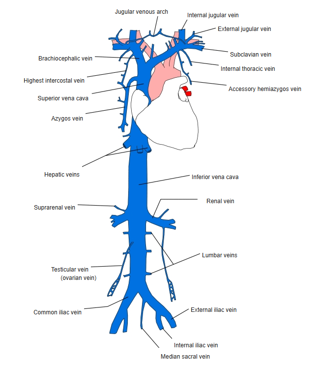
Venae cavae and their tributaries
Beginning
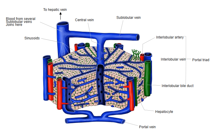
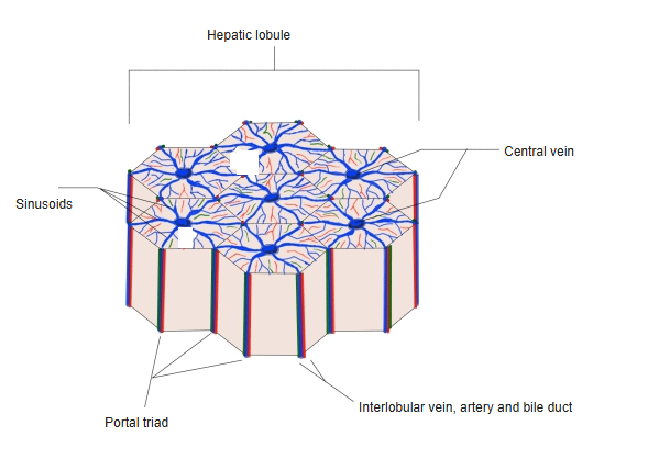
- They begin as the central veins of the hepatic lobules, which join to form sublobular veins.
Blood vessels of the hepatic lobule 
Hepatic lobule and central vein
Clinical notes
- Obstruction of the hepatic veins (Budd–Chiari syndrome) causes liver enlargement, ascites and liver damage.
References and sources
- Further reading: Hepatic veins, Wikipedia.