Latin: Arteria meningea media
Overview
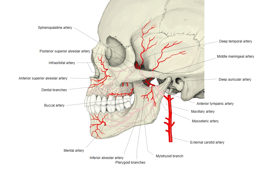
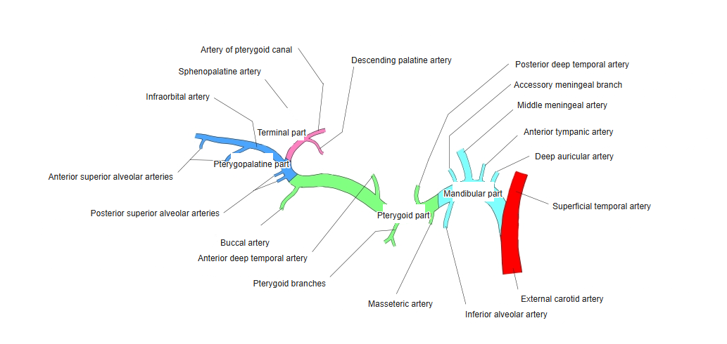
- The largest branch of the maxillary artery (first part), arising in the infratemporal fossa.
- Passes up between the two roots of the auriculotemporal nerve and enters the skull through the foramen spinosum.
- Runs in grooves on the inner surface of the skull and divides into frontal and parietal branches that supply the dura mater and the bones of the skull vault.
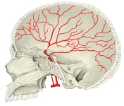
Middle meningeal artery in the skull 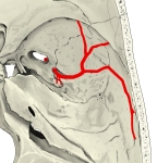
Middle meningeal artery in the cranial base
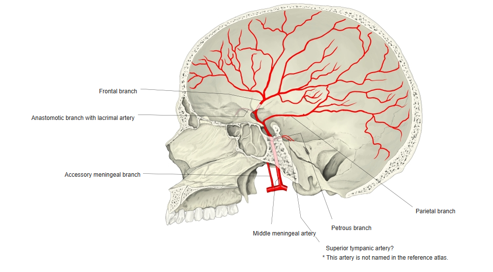
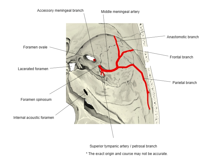
Branches
- Frontal branch and parietal branch.
- Petrosal branch (to the facial nerve) and superior tympanic artery.
- An orbital branch that joins the lacrimal artery.
Illustrations
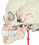
Maxillary artery and its branches (lateral view) 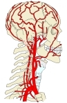
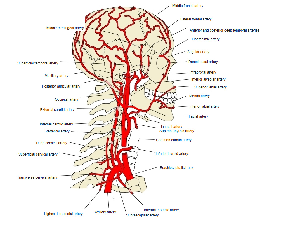
Arteries of the head and neck 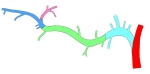
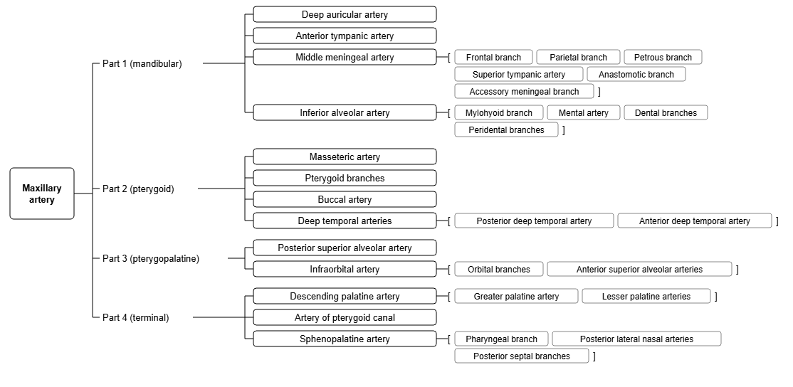
Branches by part (diagram) 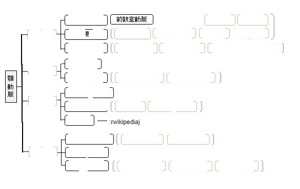
Branches of the maxillary artery (chart) 
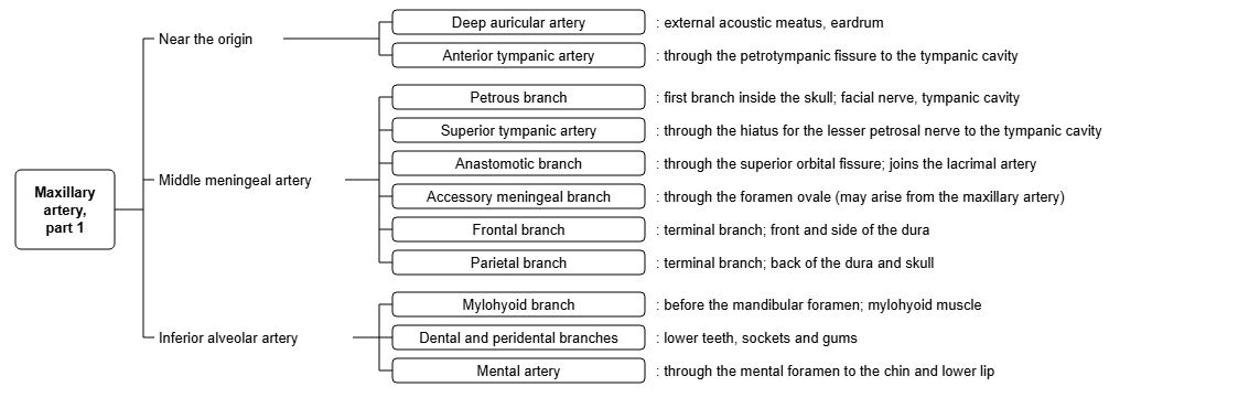
Branches of the first part of the maxillary artery (chart)
Clinical notes
- The frontal branch lies under the pterion, where the skull is thin; a blow here can tear it and cause an epidural hematoma.
References and sources
- Further reading: Middle meningeal artery, Wikipedia.