Latin: Arteria cerebri posterior
Overview
- One of the two terminal branches of the basilar artery, at the upper border of the pons.
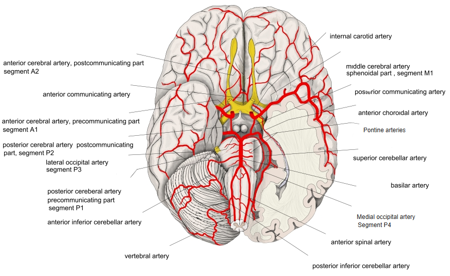
- Joined by the posterior communicating artery, then winds around the cerebral peduncle to the medial and lower surfaces of the occipital and temporal lobes.
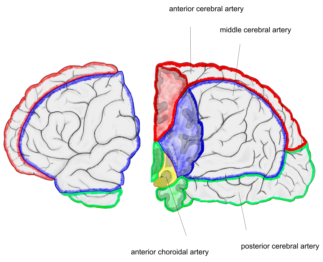
- Supplies the occipital lobe (including the visual cortex), the lower temporal lobe, the thalamus and the midbrain.
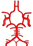
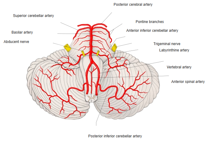
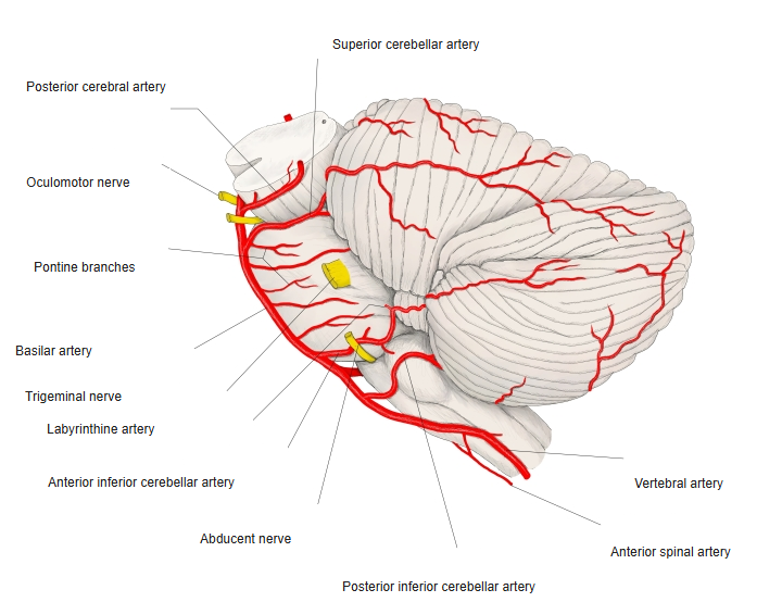
Arteries of the brainstem and cerebellum (diagram) 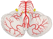
Arteries of the brainstem (front view)
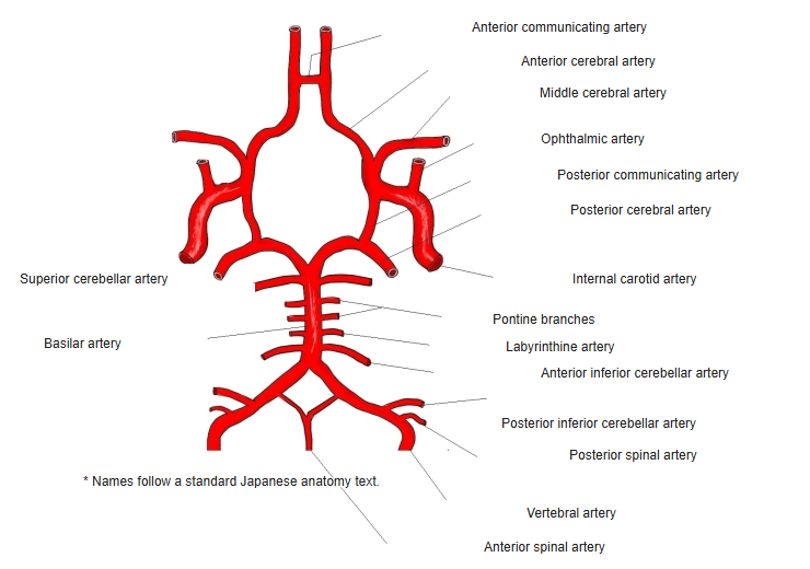
Branches
| Group | Branches |
|---|---|
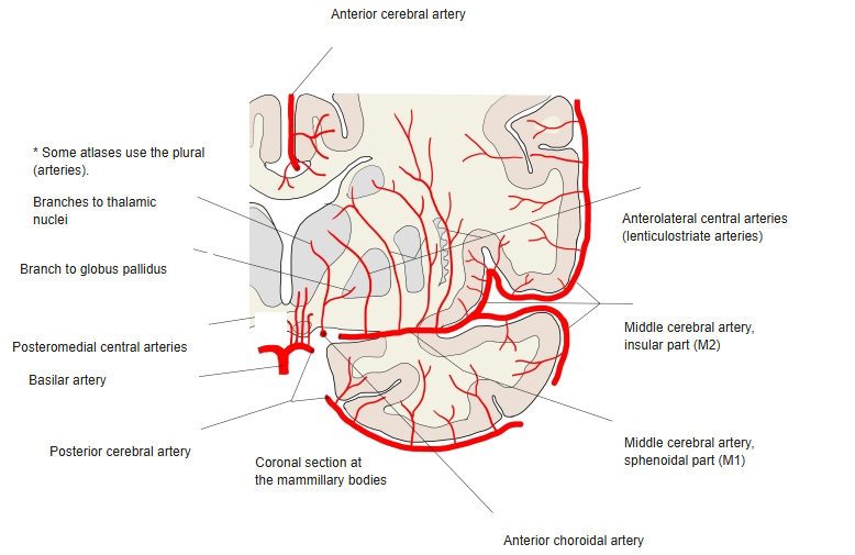
| Central branches | Posteromedial central arteries (including thalamoperforating arteries), posterolateral central arteries (thalamogeniculate arteries), peduncular branches |
| Choroid branches | Posterior medial and posterior lateral choroid branches |
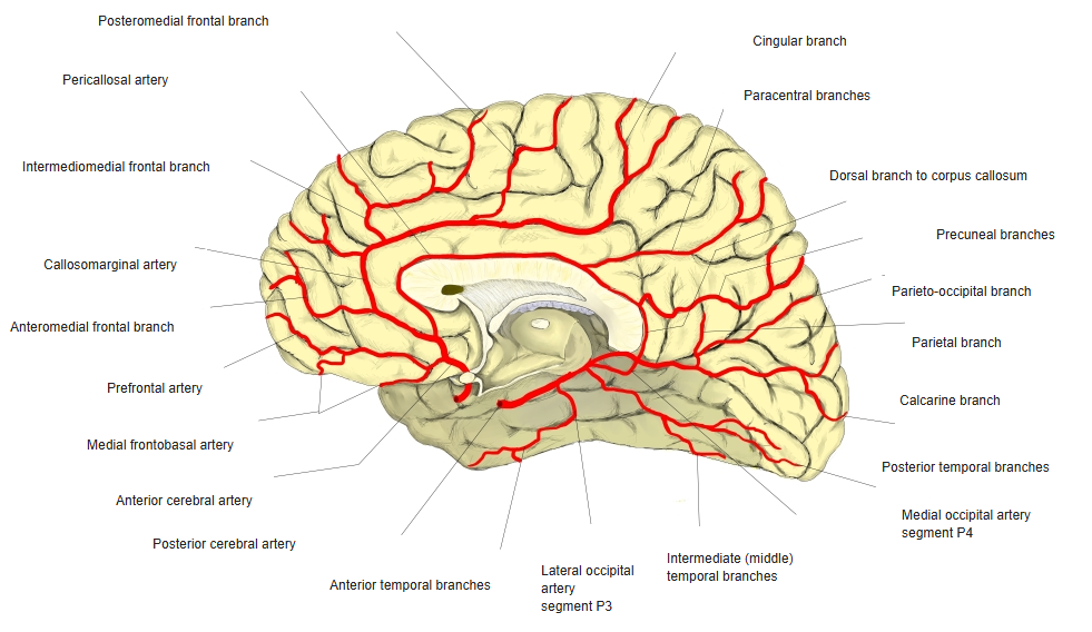
| Cortical branches | Lateral occipital artery (anterior and posterior temporal branches), medial occipital artery (calcarine, parieto-occipital and other branches) |
Illustrations
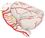
Arteries of the brainstem (side view) 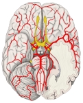
Arteries at the base of the brain 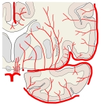
Central arteries of the brain 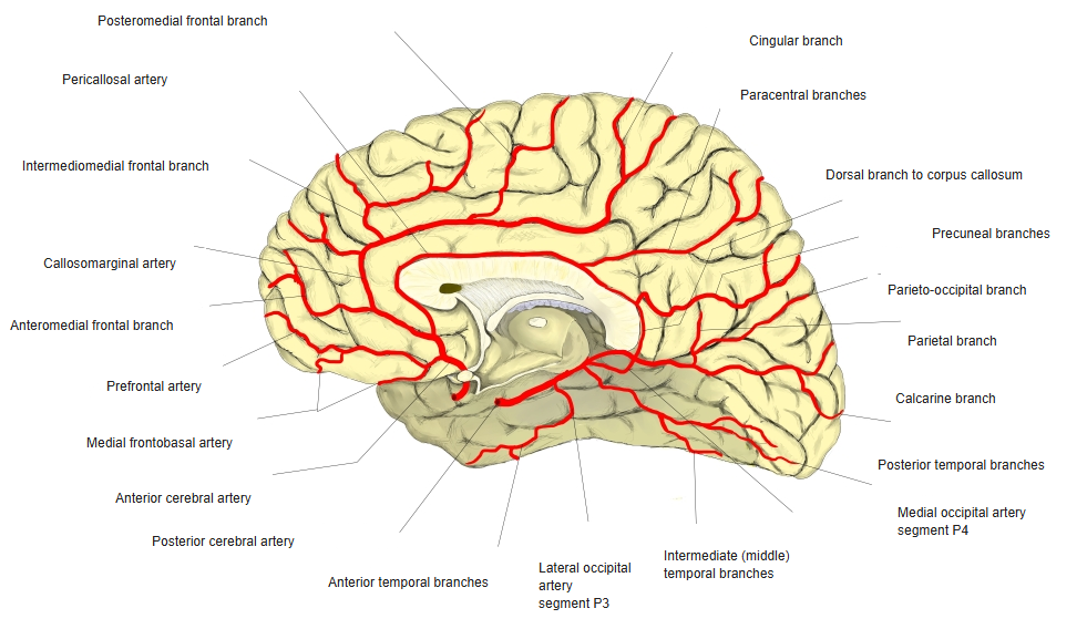
Arteries of the medial surface of the brain 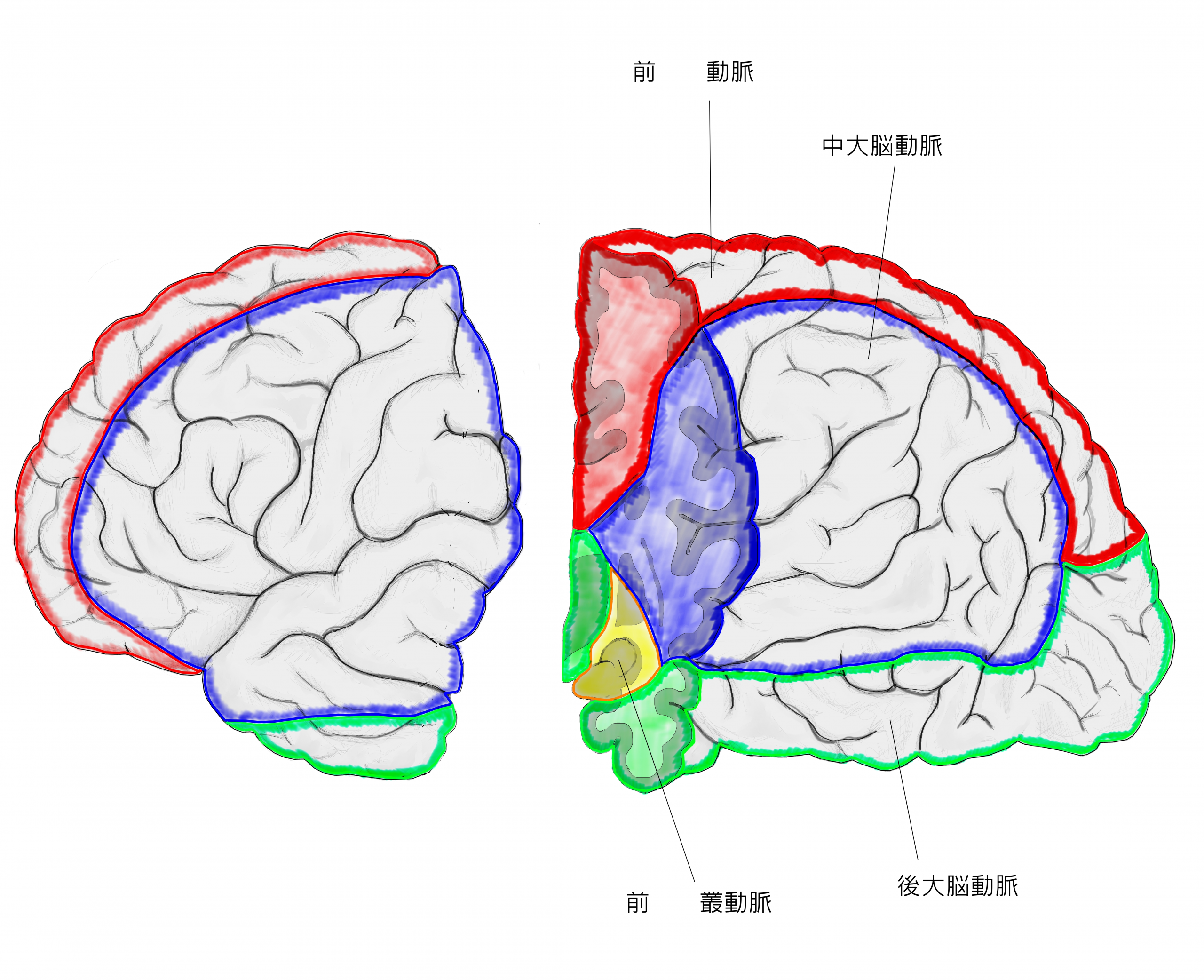
Areas supplied by the cerebral arteries
Clinical notes
- Its blockage causes loss of the opposite half of the visual field (homonymous hemianopia), often with sparing of central vision.
References and sources
- Further reading: Posterior cerebral artery, Wikipedia.