Latin: musculus fibularis tertius (peroneus tertius)
Overview
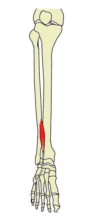
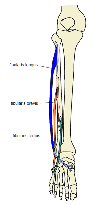
- A small muscle that separates from the lateral part of the extensor digitorum longus; it is sometimes absent.
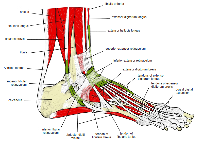
- Its tendon and those of the extensor digitorum longus are held down by a sling-like ligament between the tarsal sinus and the inferior extensor retinaculum.
- It is sometimes described as a fifth tendon of the extensor digitorum longus.
- Continuous in places with the fibularis longus and brevis, extensor hallucis longus and extensor digitorum longus.
Position in the classification
Lower Limb › Leg › Extensor group
- Tibialis anterior
- Extensor digitorum longus
- Fibularis tertius
- Extensor hallucis longus
Illustrations
Origin / insertion
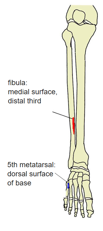
- Origin: lower third (or more) of the medial (anterior) surface of the fibula, the adjacent interosseous membrane and the intermuscular septum between it and the fibularis brevis
- Insertion: dorsal surface of the base of the 5th metatarsal
Action
- Helps to dorsiflex the foot at the ankle joint and to evert it (raises its lateral border)
Innervation
- Deep fibular nerve (L4–S1)
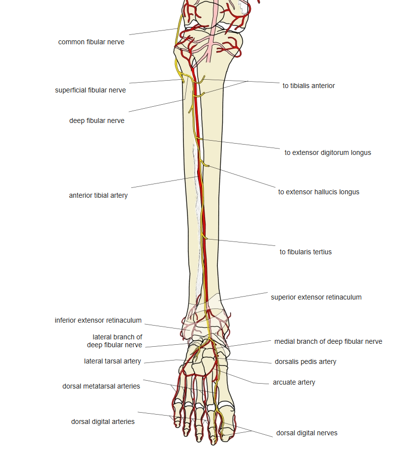
Blood supply
- Anterior tibial artery
Related terms
- Extensor digitorum longus
- Fibularis longus
- Fibularis brevis
- Fibula
- Inferior extensor retinaculum
- Deep fibular nerve
References and sources
- Further reading: Fibularis tertius, Wikipedia.