Latin: musculus extensor pollicis brevis
Overview
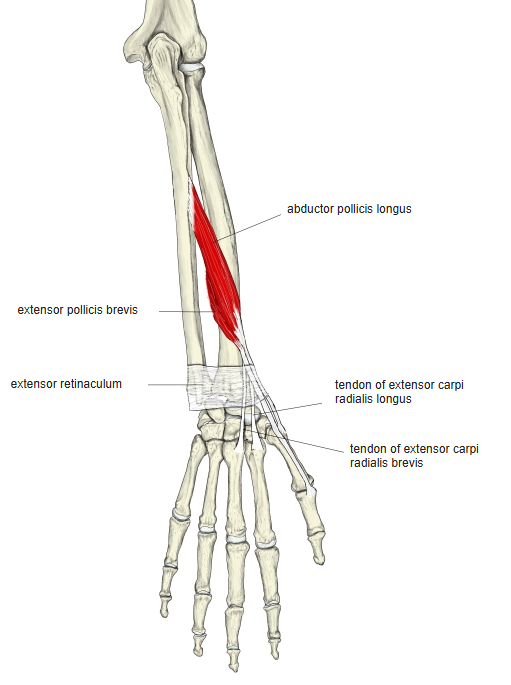
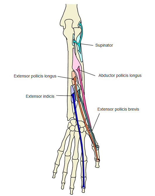
- A deep muscle on the back of the forearm, closely joined to the abductor pollicis longus; the two form one developmental and functional unit, and their bellies are often fused.
- Its tendon runs with that of the abductor pollicis longus in the same groove on the lateral side of the lower end of the radius.
- Its tendon fibres join the end of the extensor pollicis longus tendon to form a weak dorsal expansion.
- Continuous in places with the extensor pollicis longus and abductor pollicis longus.
Position in the classification
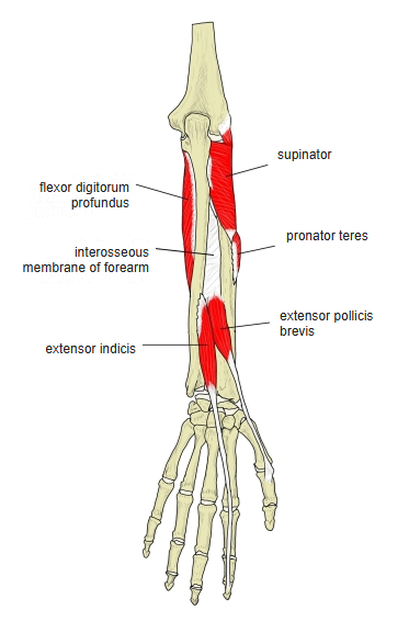
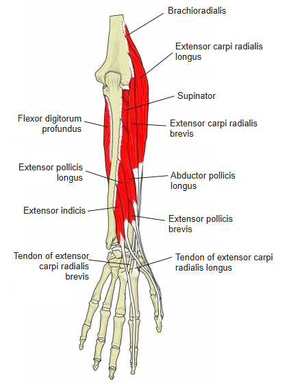
Upper Limb › Forearm › Extensor group › Deep posterior group
- Supinator
- Abductor pollicis longus
- Extensor pollicis brevis
- Extensor pollicis longus
- Extensor indicis
Illustrations
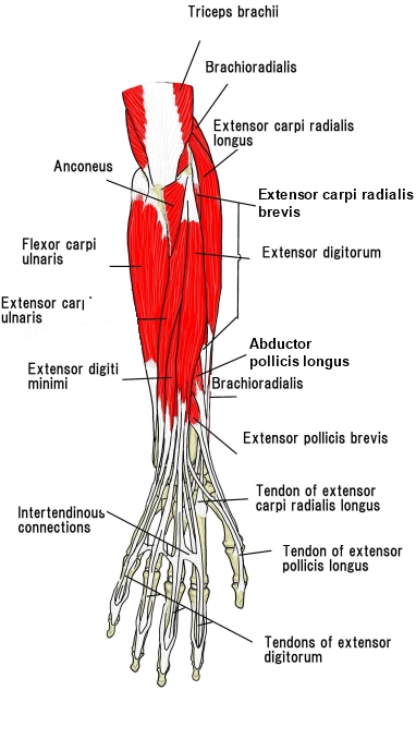
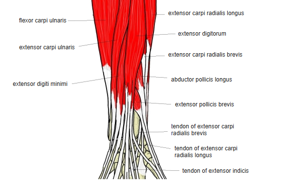
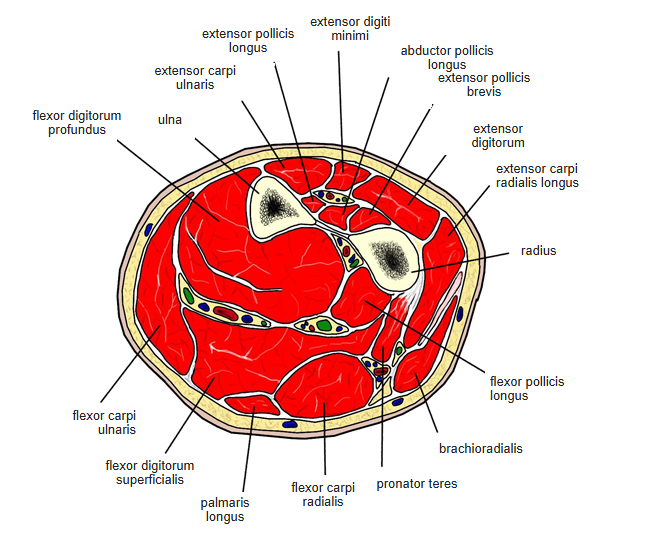
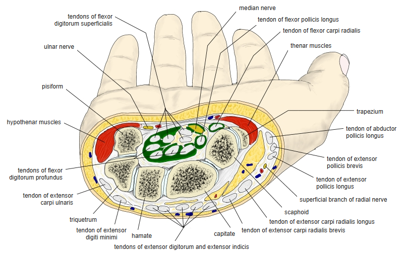
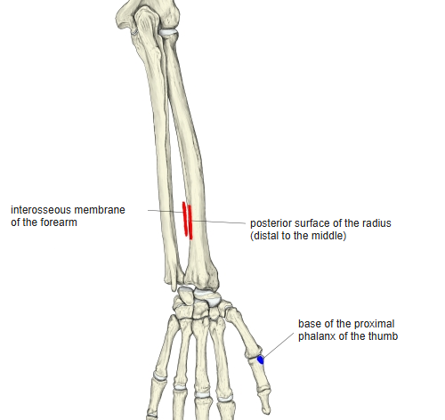
Origin / insertion
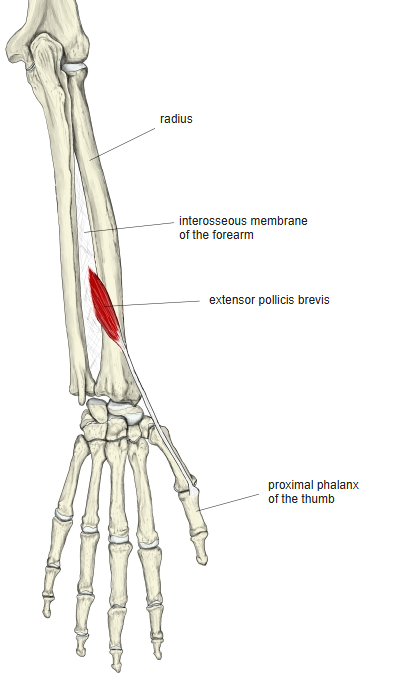
- Origin: posterior surface of the radius, distal to the abductor pollicis longus, and the interosseous membrane
- Insertion: base of the proximal phalanx of the thumb
Action
- Extends the proximal phalanx of the thumb and abducts the thumb
Innervation
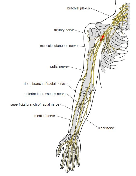
- Deep branch of the radial nerve (posterior interosseous nerve), C7–C8; some sources C6–C8
Blood supply
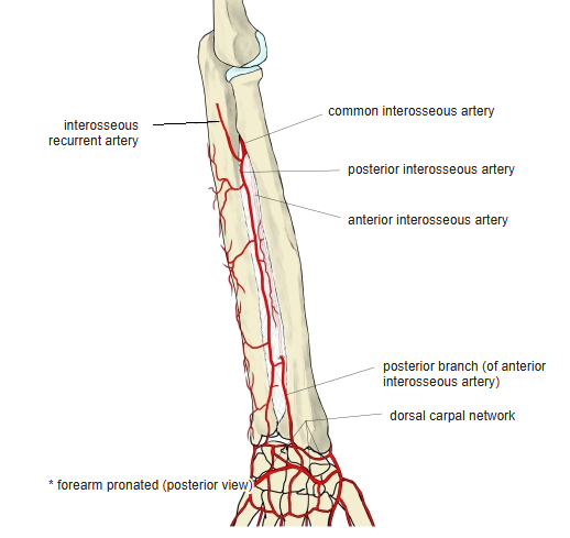
- Posterior interosseous artery
Anatomical variations
- Often completely fused with the abductor pollicis longus; its belly or tendon may be doubled
- May be absent; its tendon may fuse with that of the extensor pollicis longus or abductor pollicis longus
Related terms
- Abductor pollicis longus
- Extensor pollicis longus
- Radius
- Extensor retinaculum
- Radial nerve
References and sources
- Further reading: Extensor pollicis brevis muscle, Wikipedia.