Latin: musculus fibularis brevis (peroneus brevis)
Overview
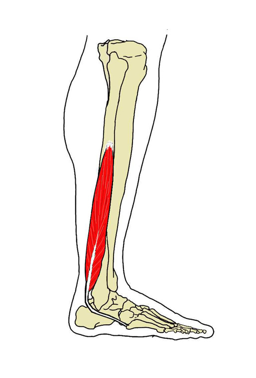
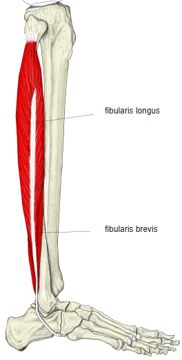
- The shorter of the two lateral leg muscles, lying under the fibularis longus.
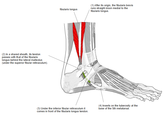
- Its fibres run vertically downward, deep (medial) to the fibularis longus.
- Behind the lateral malleolus its tendon lies in front of the fibularis longus tendon, in a common synovial sheath (under the superior fibular retinaculum); on the lateral side of the calcaneus it passes above the fibular trochlea and the fibularis longus tendon, in a separate canal under the inferior fibular retinaculum.
- The pull of its tendon can cause an avulsion fracture of the tuberosity of the 5th metatarsal (distinct from a Jones fracture, which lies slightly more distal).
- A slip of its tendon may continue to the extensor expansion of the 5th toe; in lower mammals this is a weak tendon, a remnant of a long extensor.
- Continuous in places with the fibularis longus, flexor hallucis longus, fibularis tertius and extensor digitorum longus.
Position in the classification
Lower Limb › Leg › Lateral group
- Fibularis longus
- Fibularis brevis
Illustrations
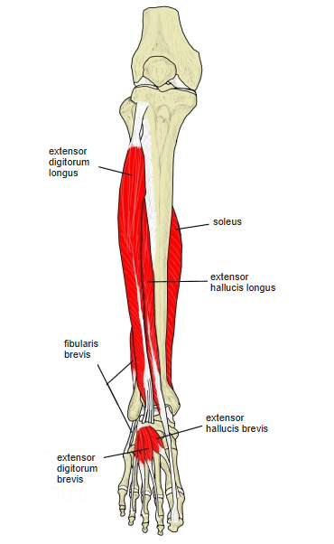
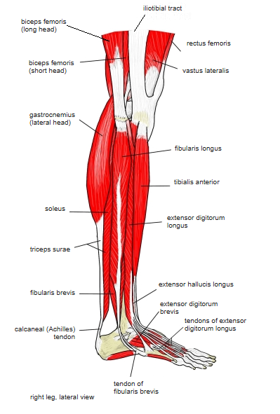
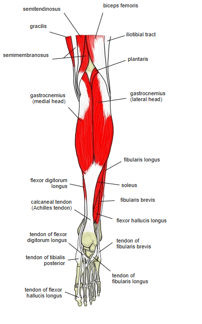
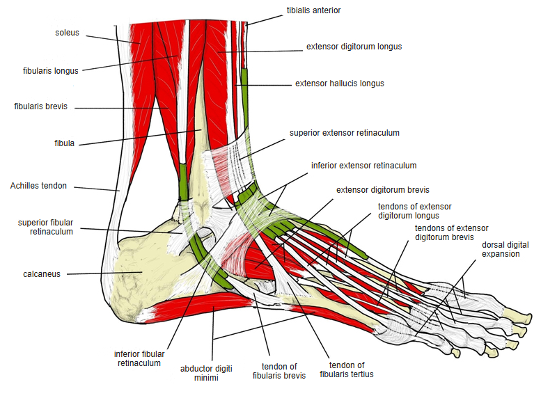
Origin / insertion
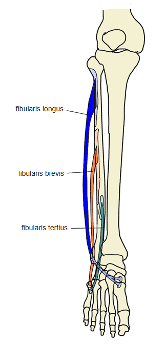
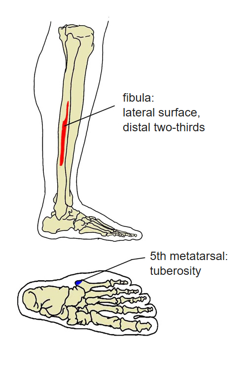
- Origin: lateral surface of the fibula, lower two-thirds; some sources add the anterior and posterior crural intermuscular septa
- Insertion: tuberosity of the 5th metatarsal
Action
- Everts the foot and steadies the ankle laterally
- Assists in plantar flexion of the foot; with the foot fixed, helps to draw the leg backwards and to steady it on the foot
Innervation
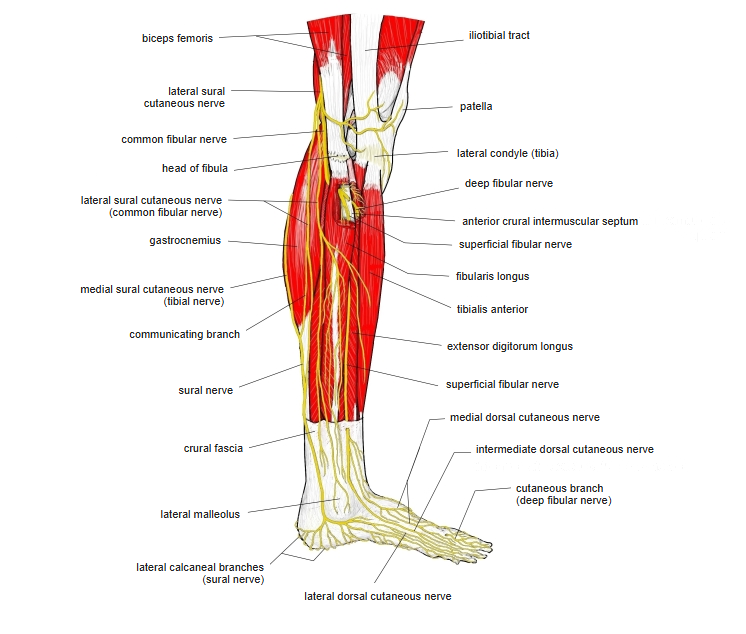
- Superficial fibular nerve (L5, S1)
Blood supply
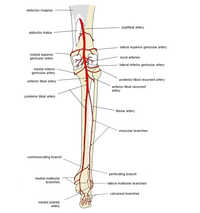
- Posterior tibial artery, fibular artery
Related terms
- Fibularis longus
- Fibularis tertius
- Fibula
- Superior fibular retinaculum
- Superficial fibular nerve
- Fibular artery
References and sources
- Further reading: Fibularis brevis, Wikipedia.