Latin: musculus fibularis longus (peroneus longus)
Overview
- The longest and most superficial of the three fibular muscles; pennate.
- Its belly lies in the upper part of the lateral side of the leg; in the lower third only its tendon continues.
- Behind the lateral malleolus it shares a synovial sheath with the fibularis brevis, held by the superior fibular retinaculum.
- It then passes below the fibular trochlea, crosses the lateral side of the cuboid and runs across the sole in a groove on the under surface of the cuboid, converted into a canal by the long plantar ligament.
- The common fibular nerve passes to the front of the leg through the gap between its attachments to the head and body of the fibula.
- Continuous in places with the fibularis brevis and tertius, extensor digitorum longus, long head of the biceps femoris, flexor hallucis longus, soleus and plantaris.
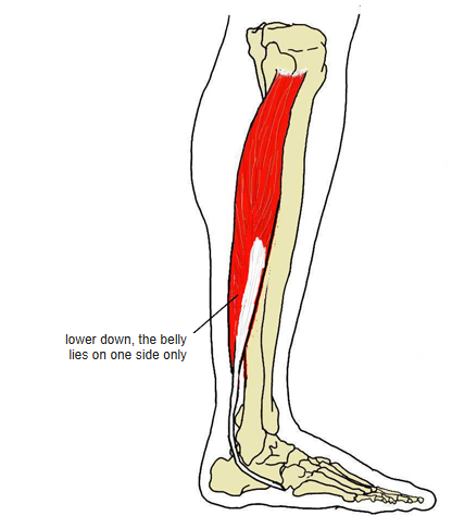
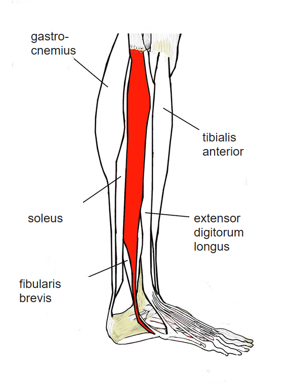
Position in the classification
Lower Limb › Leg › Lateral group
- Fibularis longus
- Fibularis brevis
Illustrations
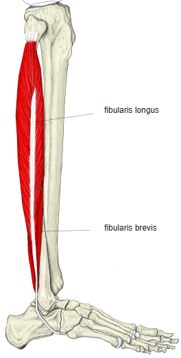
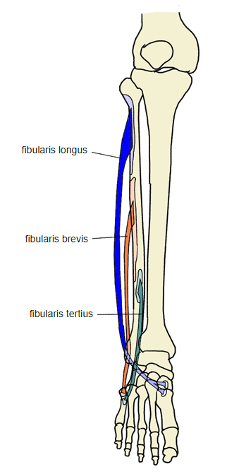
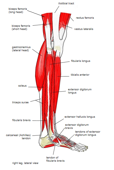
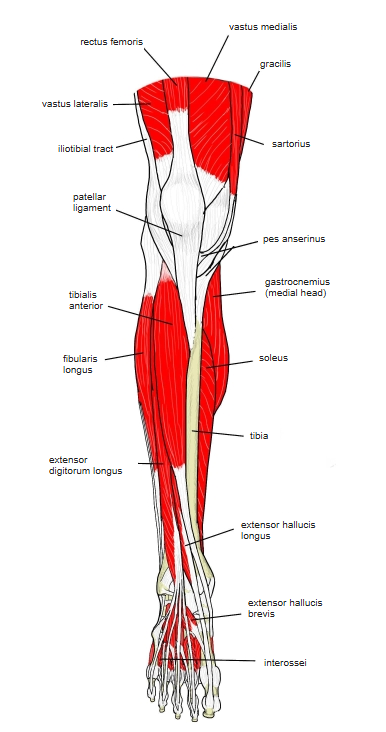
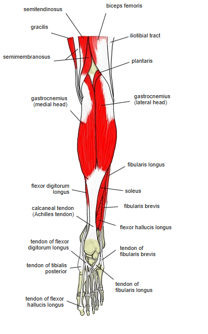
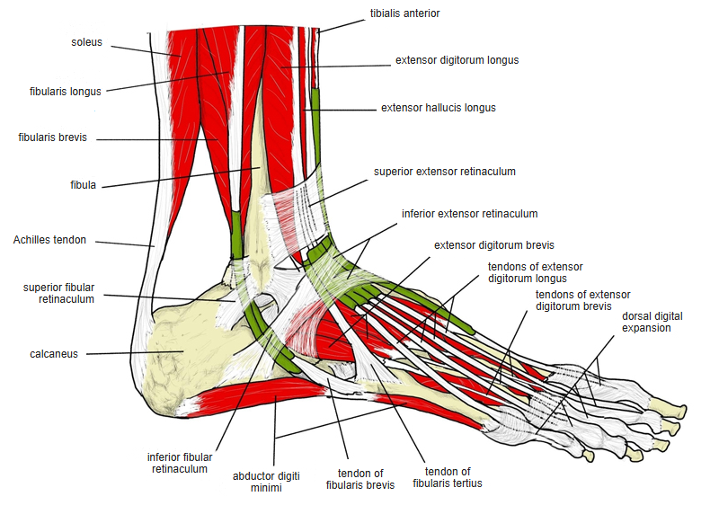
Origin / insertion
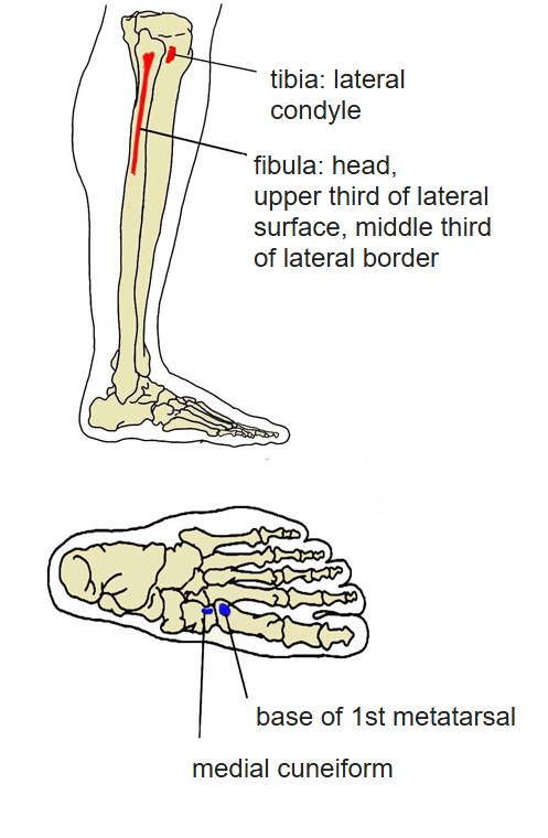
- Origin: head of the fibula and the upper two-thirds of its lateral surface, the anterior and posterior crural intermuscular septa and the crural fascia; occasionally a few fibres from the lateral condyle of the tibia (some sources also include the capsule of the tibiofibular joint)
- Insertion: medial cuneiform and base of the 1st metatarsal (sometimes also the 2nd metatarsal)
Action
- Plantar flexes and everts the foot
- With the foot fixed, helps to draw the leg backwards and to steady it on the foot (for example when standing on one leg)
Innervation
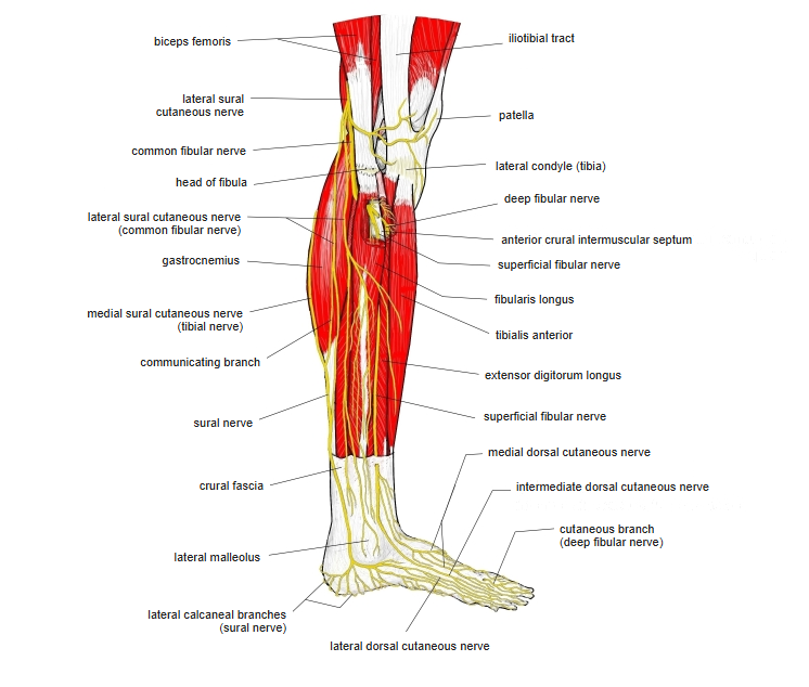
- Superficial fibular nerve (L5, S1)
Blood supply
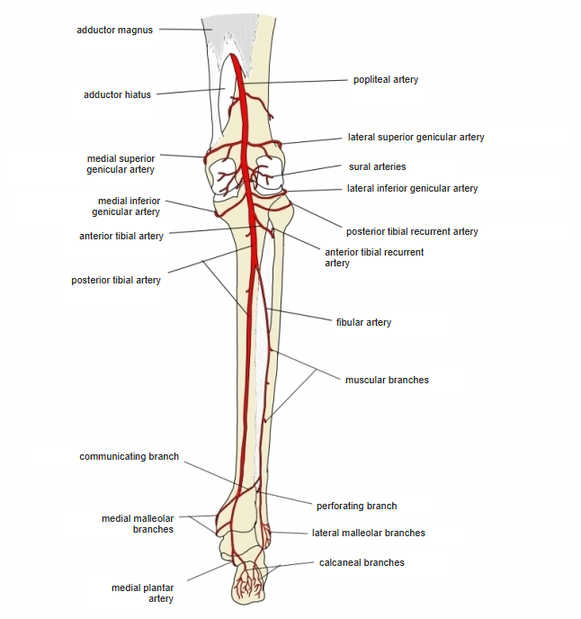
- Lateral inferior genicular artery, fibular artery, anterior tibial artery
Related terms
- Fibularis brevis
- Fibularis tertius
- Fibula
- Medial cuneiform
- Superior fibular retinaculum
- Superficial fibular nerve
- Fibular artery
References and sources
- Further reading: Fibularis longus, Wikipedia.