Major alar cartilage (Latin: cartilago alaris major)
Also called Greater alar cartilage or the lower lateral cartilage
One of the six nasal cartilages: the cartilage in the tip of the nose (nasal region). It is a thin, flexible, U-shaped plate bent on itself to support both the inner and the outer wall of the nostril on its own side. Some older texts call it the cartilage of the nasal tip, but it is not confined to the tip.
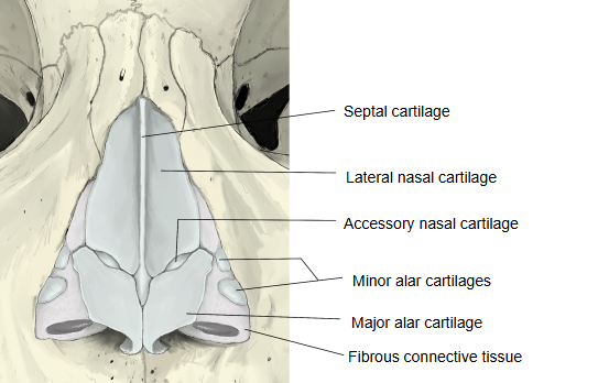
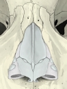 Cartilages of the nose (anterior view)
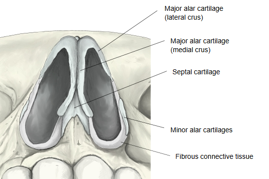
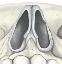 Cartilages of the nose (inferior view)
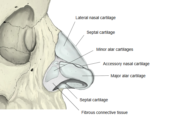
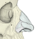 Cartilages of the nose (lateral view)
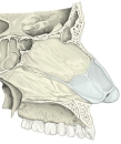 Septal cartilage (sagittal section)
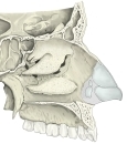 Cartilages of the side wall of the nose
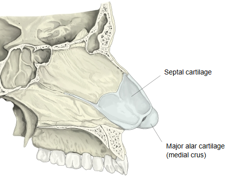
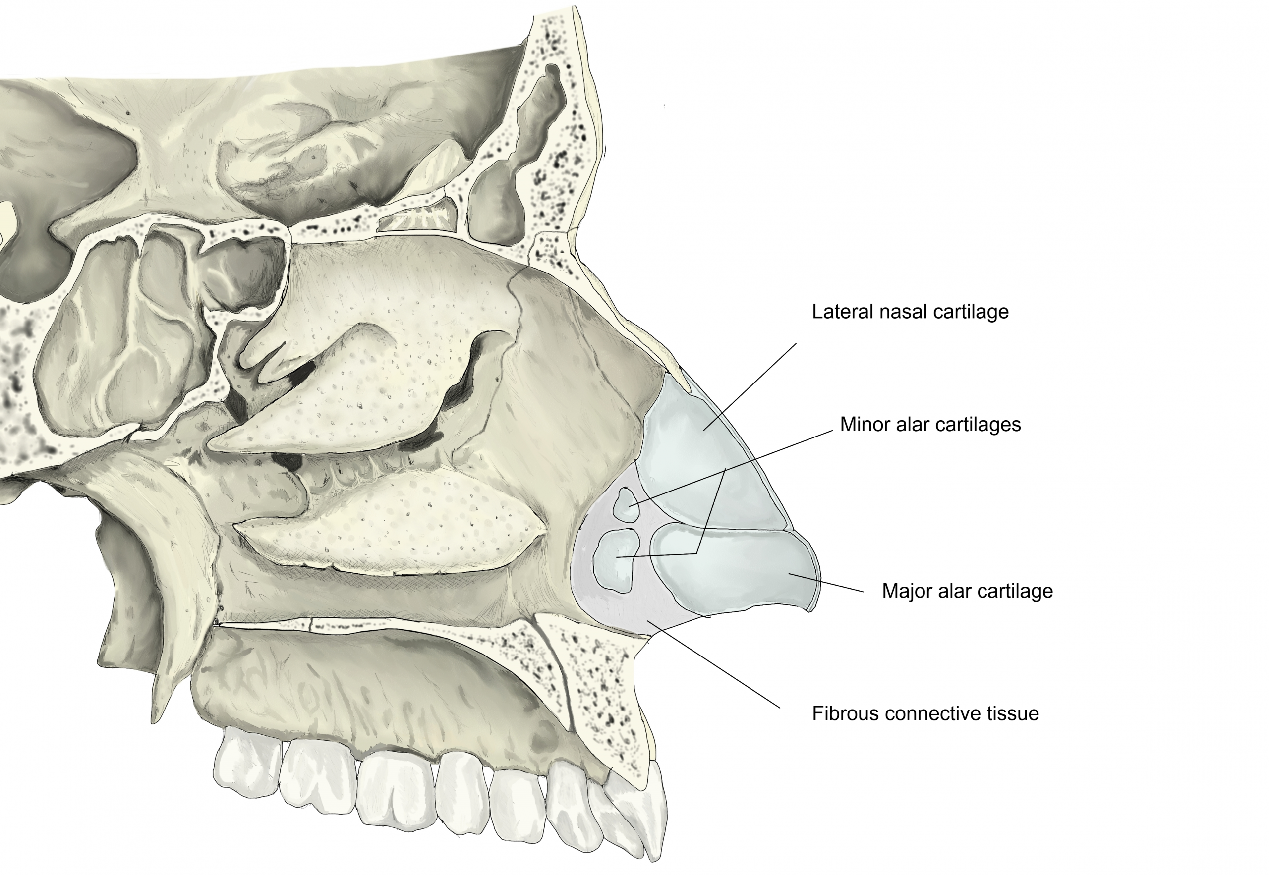
More details
· The major alar cartilage has two crura (limbs).
- Medial crus : forms the inner (medial) edge of the nostril; with the medial crus of the other side and the skin over them it forms the movable lower part of the nasal septum (columella).
- Lateral crus : curves round the ala of the nose and forms most of the outer wall of the nostril; above, it is joined to the lateral nasal cartilage by fibrous tissue.
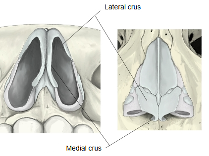
The two crura of each cartilage meet at a rounded bend (the dome) that shapes the tip of the nose, and together they surround most of the front part of the nostril and so set its shape. The right and left cartilages are separated in front by a small notch. The lateral crus is oval and flat and narrows behind, where a tough fibrous membrane containing the minor alar cartilages links it to the frontal process of the maxilla. Above, it is joined by fibrous tissue to the lateral nasal cartilage and to the front of the septal nasal cartilage. The medial crus is a narrow strip that lies loosely against the medial crus of the other side and forms part of the mobile nasal septum. The cartilage stops short of the lower edge of the ala, which is completed by fatty and fibrous tissue covered by skin; small separate pieces near the back of the lateral crus are the accessory nasal cartilages.
Related terms
- Nasal cartilages
- Minor alar cartilages
- Lateral nasal cartilage
- Septal nasal cartilage
- Accessory nasal cartilages
- Nasal septum
- Nasal vestibule
References and sources
- Further reading: Major alar cartilage, Wikipedia.
- Further reading: Nasal cartilages, Wikipedia.
- Federative International Programme for Anatomical Terminology (FIPAT). Terminologia Anatomica, 2nd ed. (TA2), 2019.