Latin: musculus piriformis

Overview

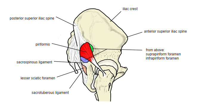
  • Leaves the pelvis through the greater sciatic foramen and divides it into the suprapiriform and infrapiriform foramina.
  • Wide at its origin, it narrows to a slender tendon; a bursa separates the tendon from the hip joint.
  • The sciatic nerve can be compressed by the muscle: piriformis syndrome.
  • Joined by connecting fibres to the gluteus maximus, medius and minimus and the superior gemellus.
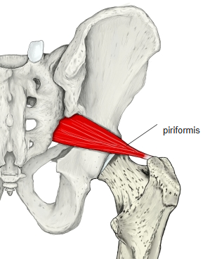
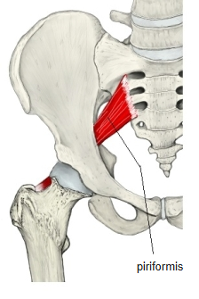
Pelvis and hip from behind with the piriformis in red.
Piriformis, posterior view
Pelvis and hip from the front with the piriformis in red.
Piriformis, anterior view

Position in the classification

Lower Limb › Hip › Outer hip muscles › Deep lateral rotators

Illustrations
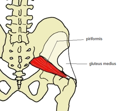
Pelvis from behind with the piriformis and the outline of the gluteus medius, labelled.
Piriformis and gluteus medius
Hip bone from the side with the piriformis and the foramina above and below it, labelled.
Piriformis and the sciatic foramina
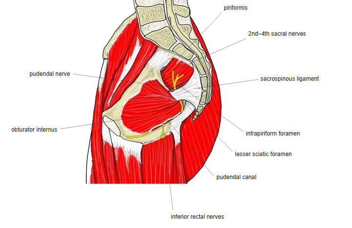
Side view of the pelvis with the piriformis, pudendal nerve and ligaments, labelled.
Piriformis and the pudendal region
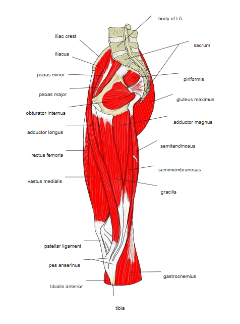
Deep muscles of the hip and thigh from behind, labelled.
Deep muscles of the hip, posterior view
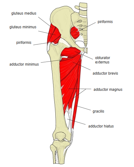
Muscles of the hip and thigh from the medial side, labelled.
Hip and thigh, medial view
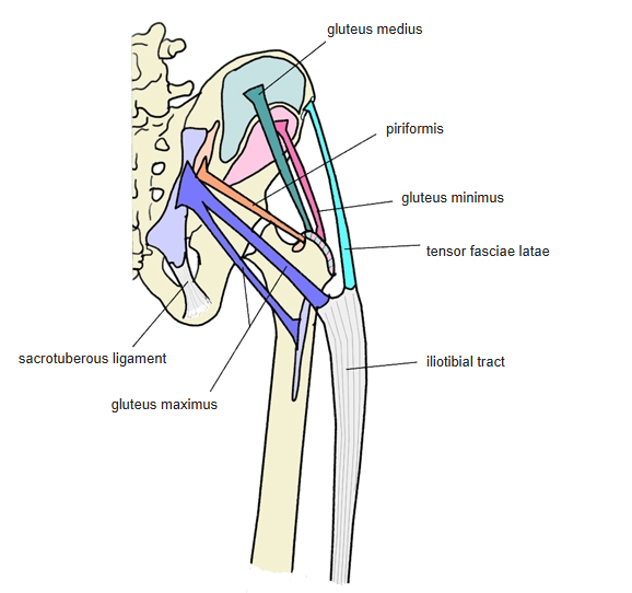
Diagram of the gluteal muscles, labelled.
Gluteal muscles, diagram
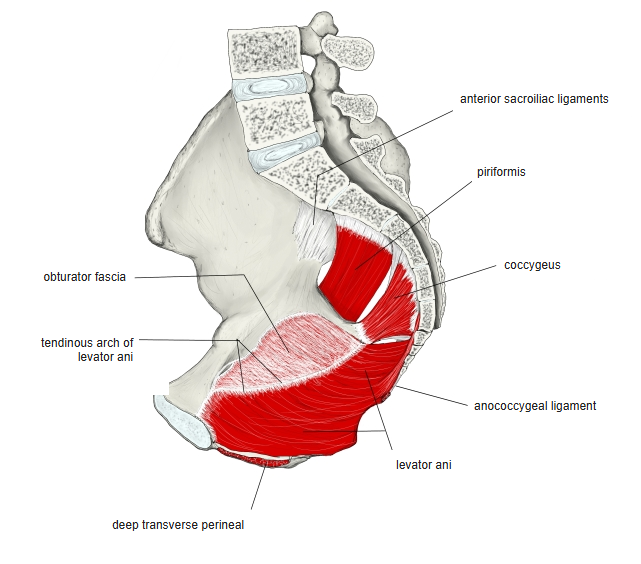
Pelvis cut in half with the piriformis, coccygeus and levator ani, labelled.
Pelvic wall, medial view
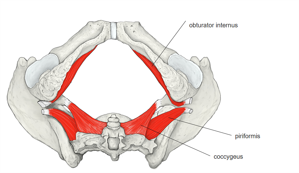
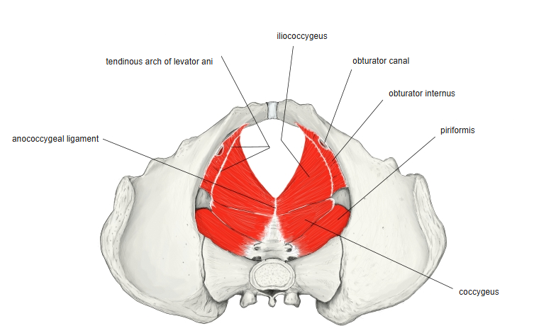
Pelvis from above with the obturator internus, piriformis and coccygeus, labelled.
Pelvic wall, superior view
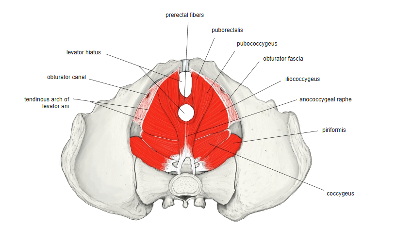
Pelvic floor from above, labelled.
Pelvic floor from above (1)
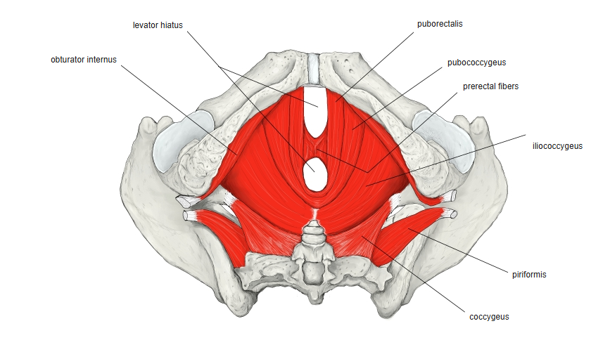
Pelvic floor from above with the parts of the levator ani, labelled.
Pelvic floor from above (2)
Pelvic floor from above, labelled.
Pelvic floor from above (3)

Origin / insertion

  • Origin: anterior (pelvic) surface of the sacrum, between and around the upper three anterior sacral foramina
  • Some sources add fibres from the capsule of the sacroiliac joint and the upper margin of the greater sciatic notch
  • Insertion: upper part of the medial surface of the greater trochanter
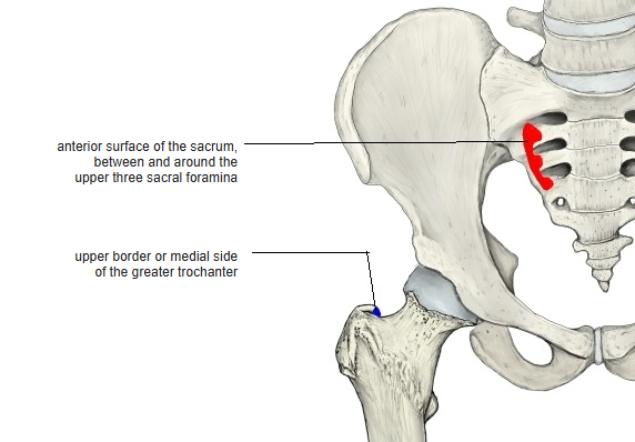
Pelvis and femur with the origin (red) and insertion (blue), labelled.
Origin and insertion

Action

  • Lateral rotation of the thigh
  • Some sources: abduction, extension and lateral rotation of the hip

Innervation

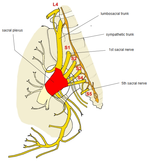
Sacral plexus from the side, labelled.
Sacral plexus

Blood supply

  • Superior gluteal artery, inferior gluteal artery
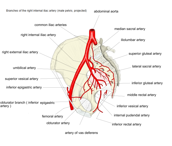
Branches of the internal iliac artery, labelled.
Internal iliac artery
  • Gluteus maximus
  • Gluteus medius
  • Gluteus minimus
  • Superior gemellus
  • Greater trochanter
  • Sacrum
  • Sciatic nerve

References and sources

  1. Further reading: Piriformis muscle, Wikipedia.