Latin: musculus latissimus dorsi
Overview
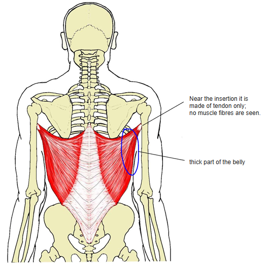
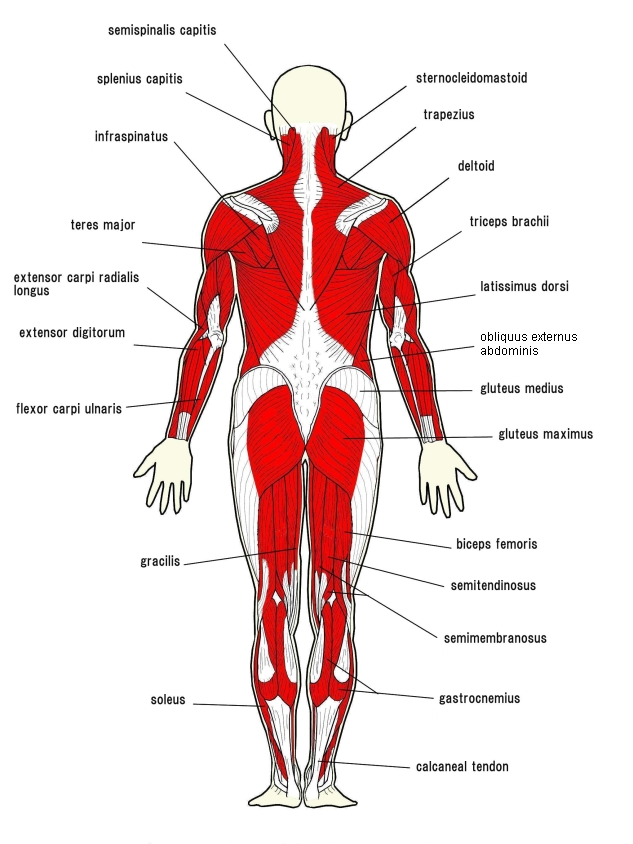
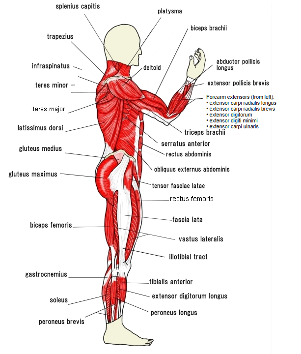
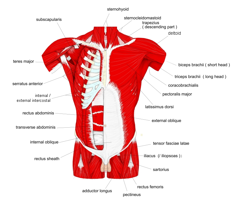
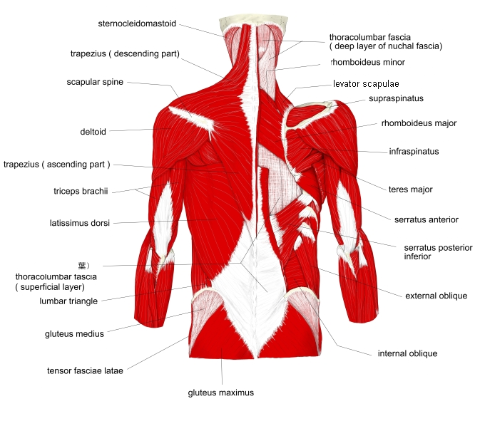
- A large, flat, triangular muscle of the lower back; a superficial back muscle of the first layer, together with the trapezius.
- Relatively thin, but thicker in its upper lateral part.
- Its upper medial part is covered by the trapezius.
- With the arm raised, its upper lateral part can be seen from the front of the body.
- It bounds two small triangles:
- Lumbar triangle (of Petit): between the latissimus dorsi, the iliac crest and the external oblique
- Triangle of auscultation: between the medial border of the scapula, the trapezius and the latissimus dorsi, which forms its lower border
Position in the classification
Illustrations
Origin / insertion
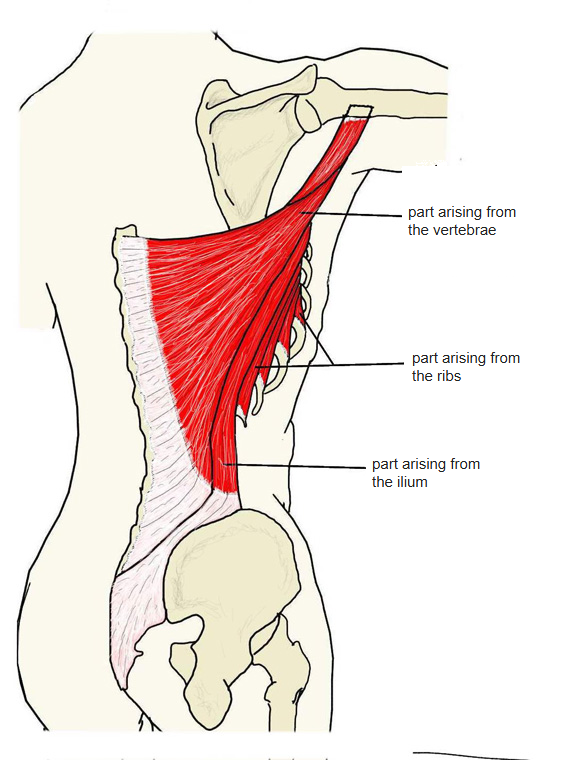
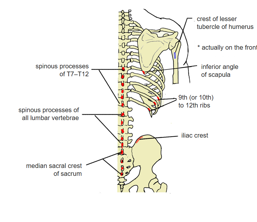
- Origin:
- Posterior layer of the thoracolumbar fascia
- Spinous processes of T7–T12 and of all lumbar vertebrae
- Median sacral crest
- Iliac crest
- 9th (or 10th) to 12th ribs
- Inferior angle of the scapula
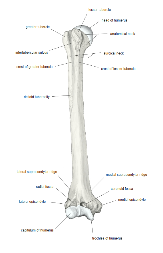
- Insertion: floor of the intertubercular sulcus (bicipital groove) of the humerus; some sources: crest of the lesser tubercle
- Near its insertion the muscle consists entirely of tendon.
Action
- At the shoulder joint: adduction, extension and medial rotation of the arm
- It also assists extension and lateral flexion of the lumbar spine, and moves the scapula (downward rotation, e.g. during a pull-up).
Innervation
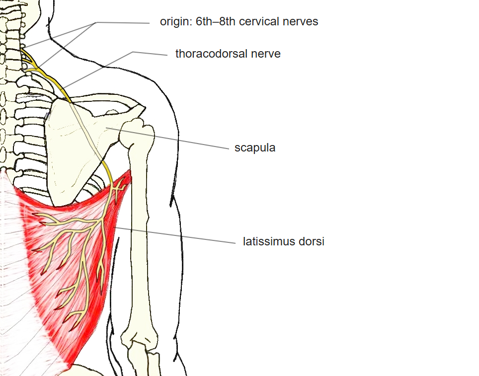
- Thoracodorsal nerve (C6–C8)
Blood supply
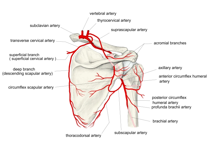
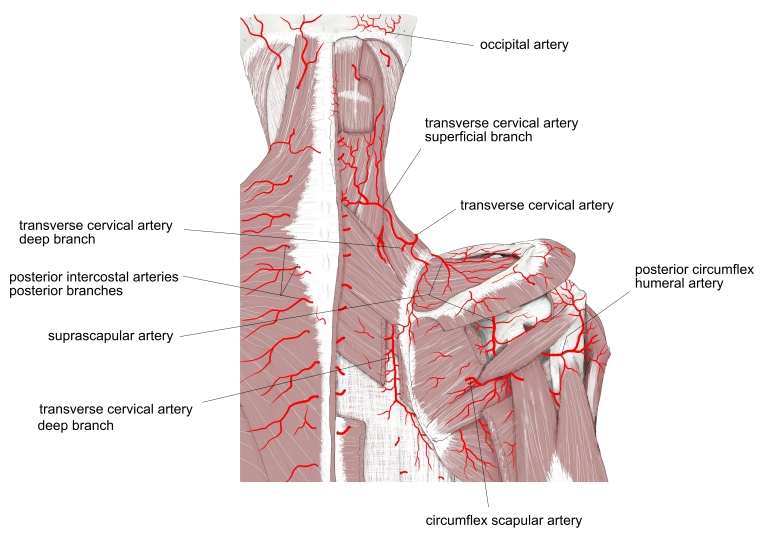
- Thoracodorsal artery (from the subscapular artery)
Anatomical variations
- May split into separate bundles; origins from the spines, iliac crest or ribs may be missing or extra
- An accessory slip from the inferior angle of the scapula is almost always present
- The tendon is often inseparable from that of the teres major
- Axillary arch (Langer's arch, 7–8%): a slip crossing the axilla, usually to the tendon of the pectoralis major
- Latissimocondyloideus (about 5%): a slip to the arm fascia, triceps or humerus
Related terms
- Superficial back muscles
- Trapezius
- Teres major
- Thoracolumbar fascia
- Lumbar triangle
- Triangle of auscultation
- Thoracodorsal nerve
References and sources
- Further reading: Latissimus dorsi muscle, Wikipedia.
- Gray H. Anatomy of the Human Body, 20th ed. (1918), “The Muscles Connecting the Upper Extremity to the Vertebral Column”, Bartleby.com.