Latin: musculus omohyoideus
Overview
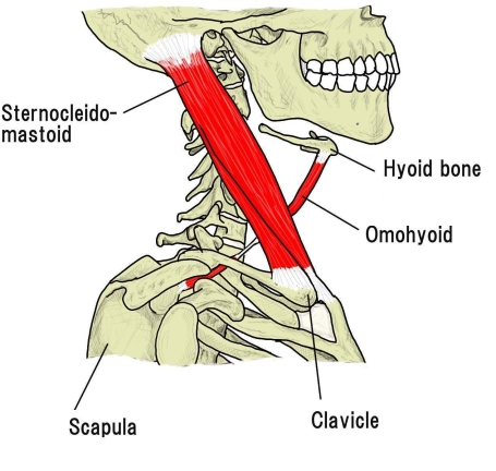
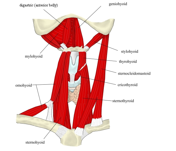
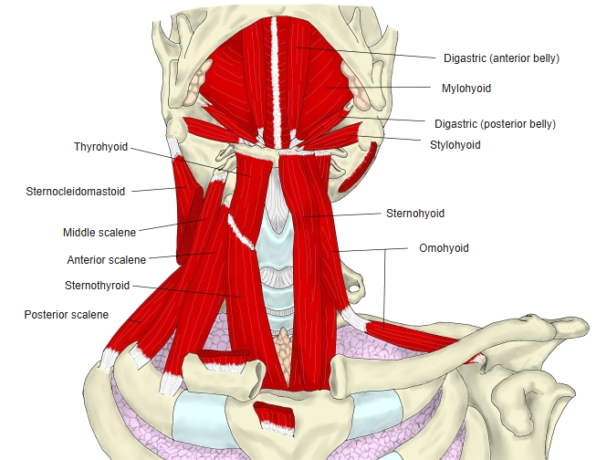
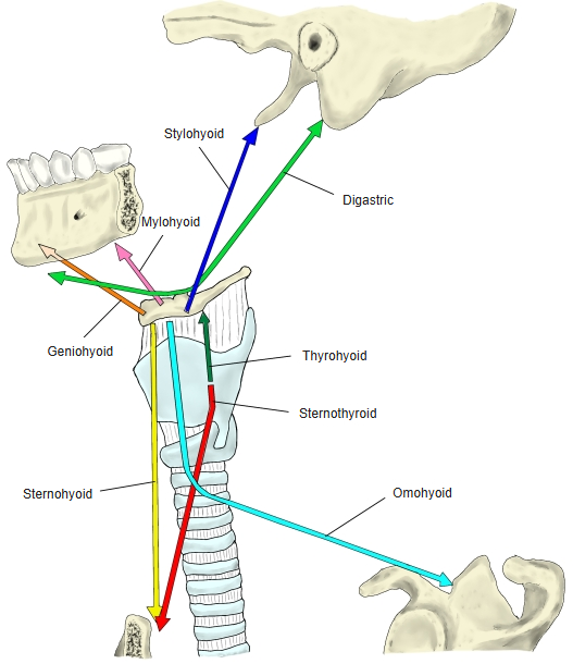
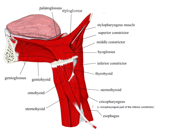
- One of the infrahyoid muscles: a thin muscle stretched between the scapula and the hyoid bone.
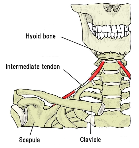
- It has two bellies, inferior and superior, joined by an intermediate tendon; as a whole the muscle forms a curve.
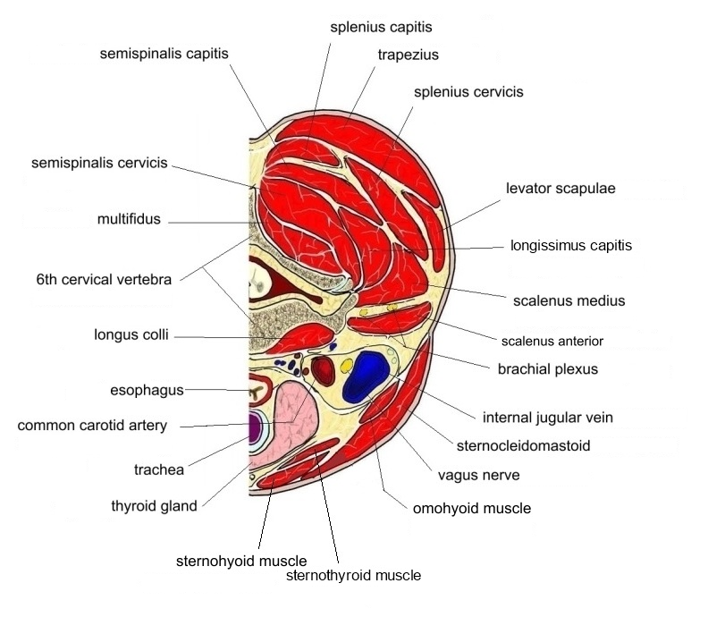
- It lies in the same layer as the sternohyoid and crosses behind the sternocleidomastoid.
- With the sternocleidomastoid it bounds the carotid triangle (above, superior belly) and the omoclavicular triangle (below, inferior belly).
Position in the classification
Neck › Superficial muscles of the neck › Infrahyoid muscles
Illustrations
Origin / insertion
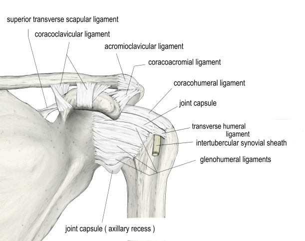
- Origin (inferior belly): superior border of the scapula near the coracoid process, and the superior transverse scapular ligament
- The inferior belly runs obliquely upward and medially, passes behind the sternocleidomastoid and becomes the intermediate tendon.
- Insertion (superior belly): lower border of the body of the hyoid bone, lateral to the sternohyoid
- The intermediate tendon is held down toward the clavicle by the deep cervical fascia and lies over the internal jugular vein.
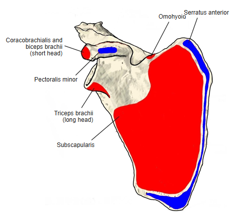
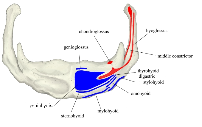
Action
- Pulls the hyoid bone downward and backward
- At the same time tenses the cervical fascia
Innervation
- Superior root of the ansa cervicalis (C1–C3)
- Some sources give C2–C3.
Blood supply
- Branches of the inferior thyroid artery
Compiled from standard anatomy references.
Anatomical variations
- May be doubled or absent
- Origin from the clavicle
- Absence or doubling of either belly
Related terms
- Infrahyoid muscles
- Sternohyoid
- Sternocleidomastoid
- Intermediate tendon
- Superior transverse scapular ligament
- Carotid triangle
- Omoclavicular triangle
- Hyoid bone
- Ansa cervicalis
References and sources
- Further reading: Omohyoid muscle, Wikipedia — also the source for the blood supply.