Latin: musculus extensor indicis
Overview
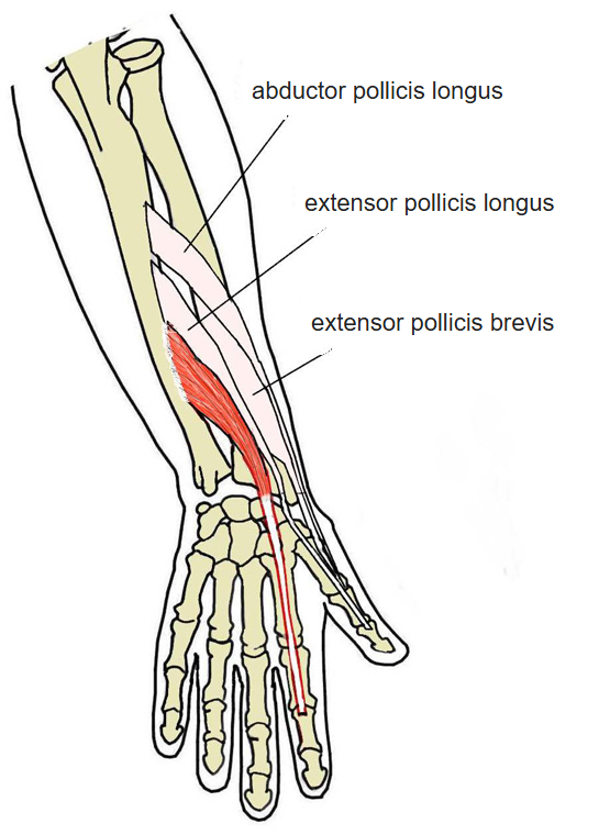
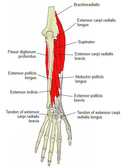
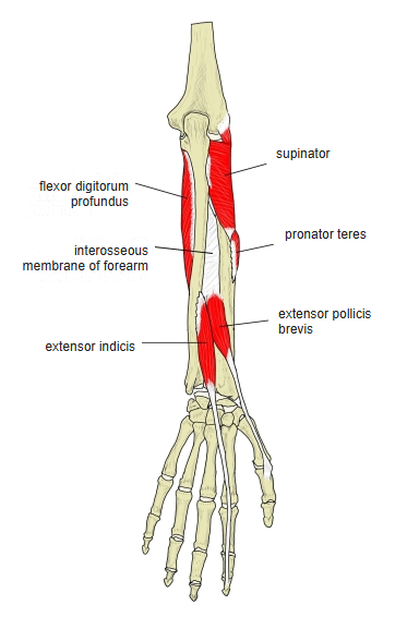
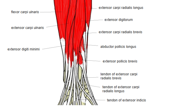
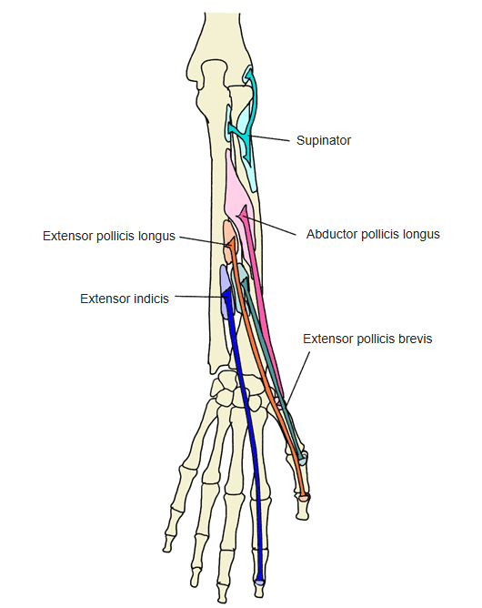
- One of the 11 muscles of the extensor group of the forearm, in the deep layer (with the supinator, abductor pollicis longus and the long and short extensors of the thumb).
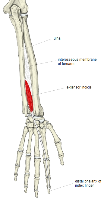
- A narrow, elongated muscle running down the back of the forearm on the ulnar side of, and parallel to, the extensor pollicis longus.
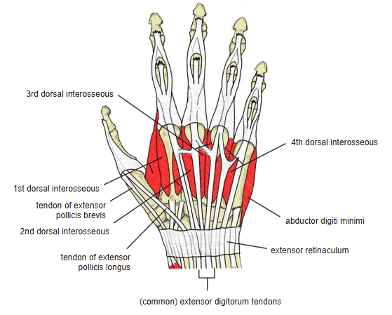
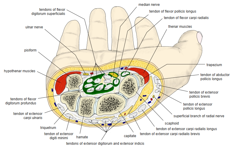
- Its tendon passes under the extensor retinaculum through the 4th tendon compartment, together with the extensor digitorum, and reaches the index finger.
- Part of the muscle belly lies just under the skin between the extensor digiti minimi and the ulna in the distal forearm; its pennate fibres reach as far as the dorsum of the wrist.
- Continuous in places with the extensor pollicis longus, extensor carpi radialis brevis and extensor digitorum.
Position in the classification
Upper Limb › Forearm › Extensor group › Deep posterior group
Illustrations
Origin / insertion
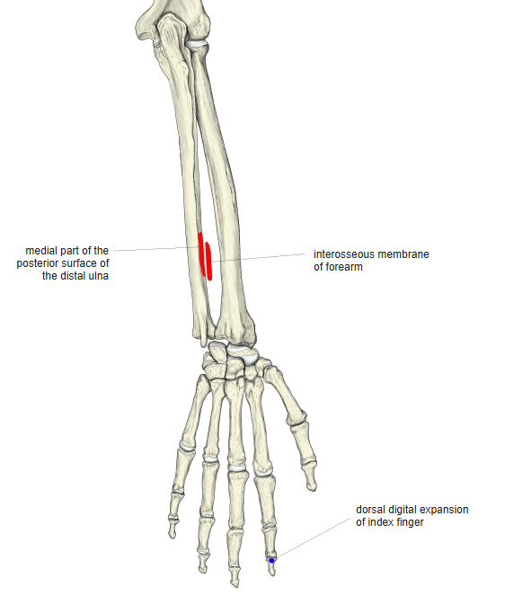
- Origin:
- Posterior surface of the distal third of the ulna
- Interosseous membrane of the forearm
- Fascia of the extensor carpi ulnaris
- Insertion: dorsal digital expansion of the index finger, together with the index-finger tendon of the extensor digitorum
- Opposite the head of the 2nd metacarpal it joins the ulnar side of that extensor digitorum tendon.
- Unlike the extensor digitorum tendons, it has no juncturae tendinum linking it to neighbouring tendons.
Action
- Extends the index finger
- Assists in extending the wrist
- Because the index and little fingers each have an extra extensor, they can be moved more independently than the other fingers.
Innervation
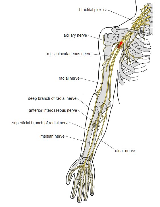
- Posterior interosseous nerve, a branch of the radial nerve (C7–C8; some sources C6–C8)
Blood supply
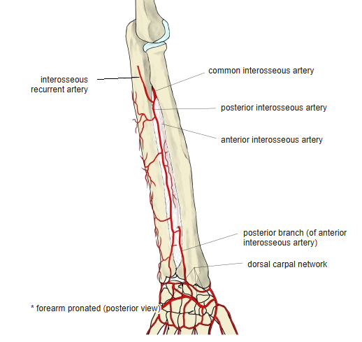
- Posterior interosseous artery
- Perforating branch of the anterior interosseous artery
Anatomical variations
- Rarely absent or doubled
- May have two tendons to the index finger, or send one tendon to the middle or ring finger
- May insert on the radial side of the extensor digitorum tendon (extensor indicis radialis), or on both sides
- Extra hand extensors such as the extensor medii proprius are regarded as related variants
Related terms
- Extensor digitorum
- Extensor pollicis longus
- Extensor digiti minimi
- Ulna
- Interosseous membrane of forearm
- Dorsal digital expansion
- Extensor retinaculum
- Juncturae tendinum
- Radial nerve
- Posterior interosseous nerve
- Posterior interosseous artery
- Anterior interosseous artery
References and sources
- Further reading: Extensor indicis muscle, Wikipedia.