Latin: Arteria centralis retinae
Overview
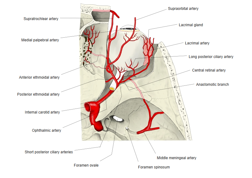
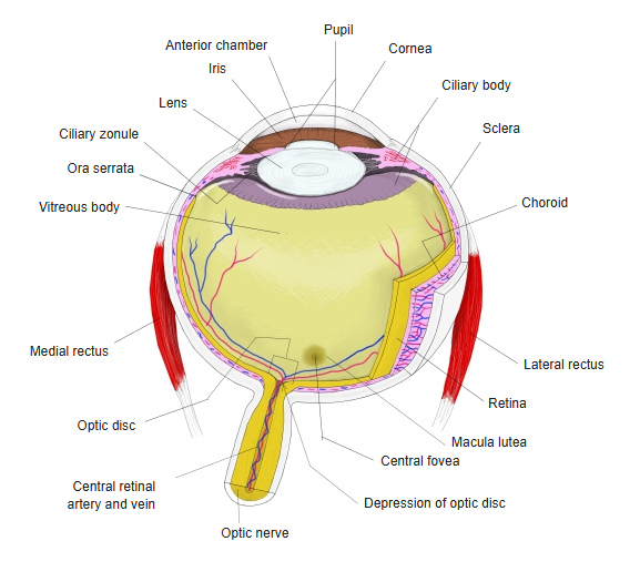
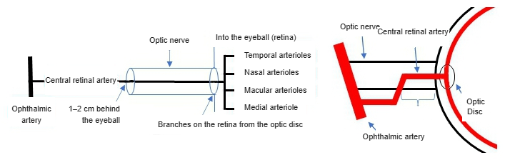
- A small branch of the ophthalmic artery that enters the optic nerve about 1 cm behind the eyeball and runs in its centre to the optic disc.
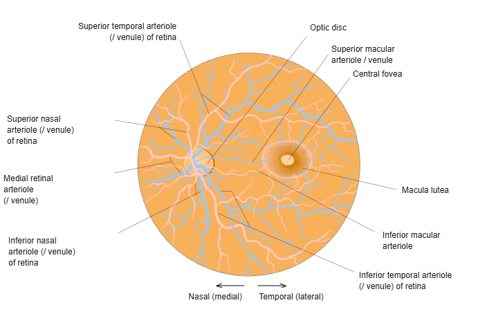
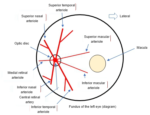
- Divides on the retina into superior and inferior branches, each with nasal and temporal arterioles; small macular and medial arterioles are also named.
- It is an end artery: it supplies the inner layers of the retina with no effective anastomoses.
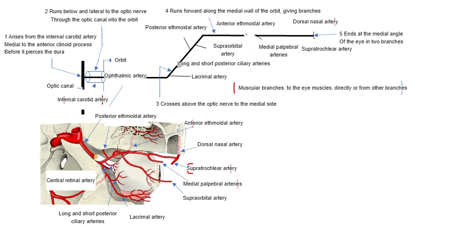
Ophthalmic artery and its branches 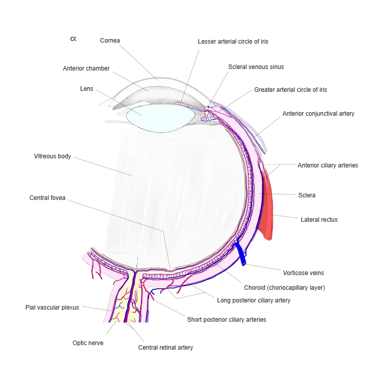
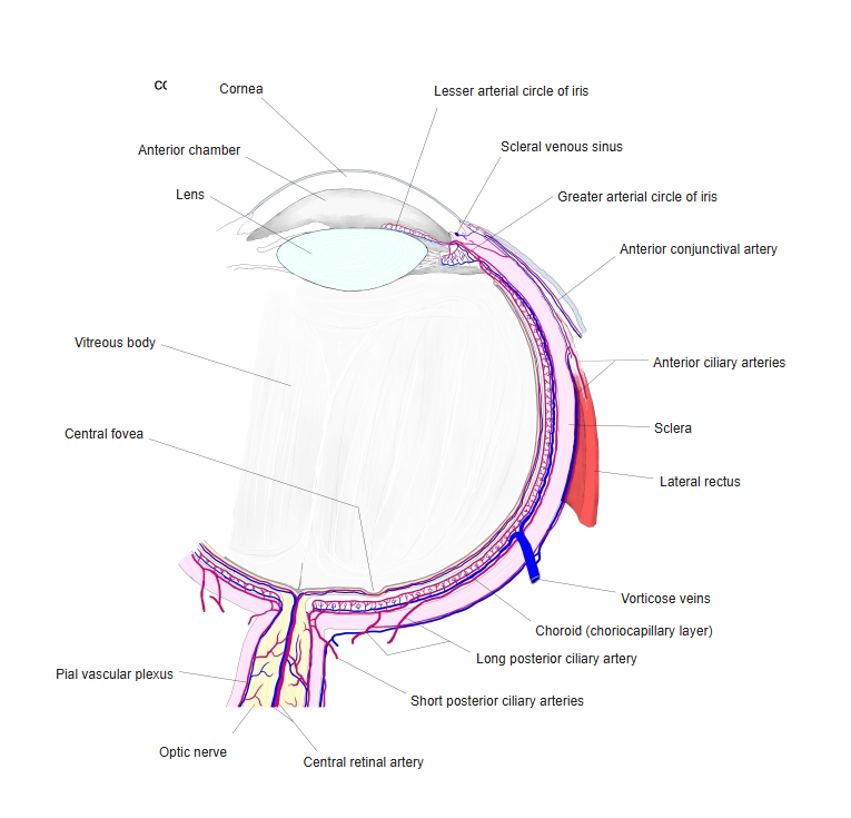
Blood supply of the eyeball
Illustrations
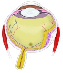
Structure of the eyeball 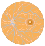
Arterioles of the retina 
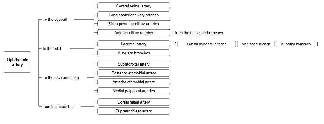
Branches of the ophthalmic artery (chart) 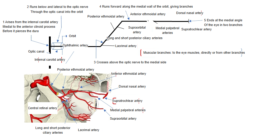
Course and branches of the ophthalmic artery 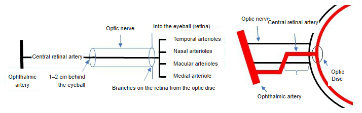
Course of the central retinal artery (diagram) 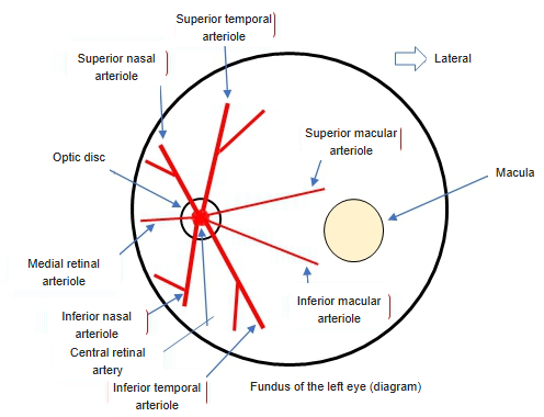
Arterioles of the retina (diagram)
Clinical notes
- Blockage (central retinal artery occlusion) causes sudden painless loss of vision in one eye.
- Its branches can be seen directly with an ophthalmoscope.
References and sources
- Further reading: Central retinal artery, Wikipedia.