Latin: Venae meningeae mediae
Overview
- Paired veins that accompany the middle meningeal artery in grooves on the inner surface of the skull.
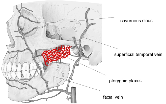
- Leave the skull through the foramen spinosum (or the foramen ovale) and end in the pterygoid plexus.
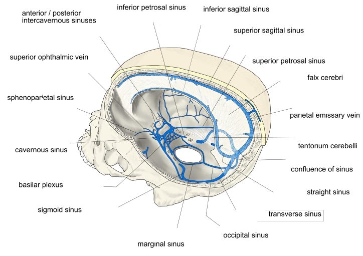
- Some branches end in the superior sagittal sinus or the sphenoparietal sinus.
Pterygoid plexus 
Dural venous sinuses
Clinical notes
- Lie between the artery and the bone, so they may be torn in skull fractures and contribute to epidural bleeding.
References and sources
- Further reading: Middle meningeal veins, Wikipedia.