Costal cartilage (Latin: cartilago costalis)
| 1 | Costal cartilages 1–7 | · Join the costal notches on the lateral border of the sternum. · They become longer from above downwards. | 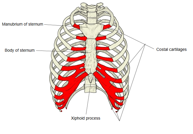 |
| 2 | Costal cartilages 8–10 | · Each joins the lower border of the cartilage above (the 8th joins the 7th); together with the 7th they form the costal arch. | |
| 3 | Costal cartilages 11 and 12 | · Short, pointed tips that end free among the muscles of the abdominal wall. |
The costal cartilages of the upper seven pairs of ribs are bars of hyaline cartilage that join the ribs to the sides of the sternum; these joints are strengthened by the radiate sternocostal ligaments, and the first cartilage is also tied to the clavicle by the costoclavicular ligament. Cartilages 8–10 each join the cartilage above, and those of the floating ribs (11 and 12) end in the muscles of the abdominal wall. Thanks to the costal cartilages the thoracic wall is elastic and mobile, so the ribs can move in breathing. In old age the cartilages become harder because they calcify, starting at the surface.
Reference websites
[Wikipedia]
· Costal cartilage, Wikipedia.
· Rib, Wikipedia.
[Websites with illustrations]
· Costal cartilages (shown in red), front view (Wikimedia Commons)

· Sternocostal and interchondral joints, front view (Gray's Anatomy) (Wikimedia Commons)

· The ribs and rib cage (TeachMeAnatomy)
· Ribs: anatomy, ligaments and clinical notes (Kenhub)
Where it is found
Related terms
References and sources
- Further reading: Costal cartilage, Wikipedia.
- Further reading: Rib, Wikipedia.
- Federative International Programme for Anatomical Terminology (FIPAT). Terminologia Anatomica, 2nd ed. (TA2), 2019.