Sinusoid (Latin: vas sinusoideum)
Also called Sinusoidal capillary or the discontinuous capillary
· Also called the sinusoidal capillary or discontinuous capillary.
· Some anatomy textbooks count it among the "special capillaries".
Such textbooks divide capillaries broadly into a continuous and a fenestrated type, and name the following three special capillaries. Most English histology texts instead list three types: continuous, fenestrated and sinusoidal (discontinuous).
laries
- Fenestrated type : the endothelial cells have many small pores (fenestrae); for example in the kidney and the intestinal villi. Substances pass through the wall easily.
- Continuous type : no pores in the endothelium; for example in the brain and skeletal muscle.
capillaries
- Sinusoid : wider than ordinary capillaries, with gaps in the wall (counted as fenestrated in some texts, discontinuous in most); found in the liver and in endocrine organs such as the anterior pituitary and the adrenal gland.
- Glomerular capillaries : one artery suddenly branches into a tuft of capillaries, which join again into one artery: a "wonder net" (rete mirabile), found in the glomerulus of the kidney.
- Splenic sinus : in the spleen; long, rod-shaped endothelial cells with slits between them.
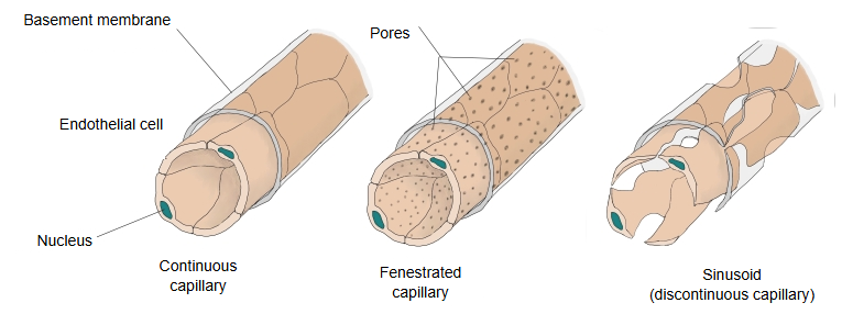 Three types of capillary
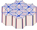 Hepatic lobules
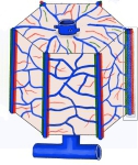 A single hepatic lobule
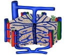 Hepatic lobule (cut-away)
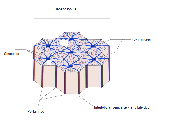
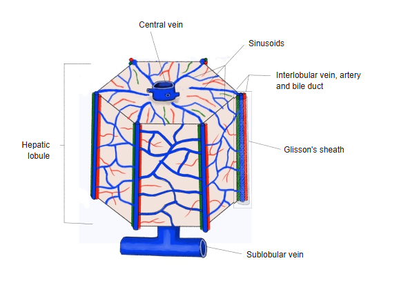
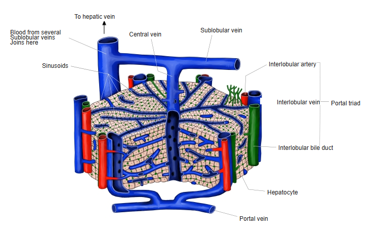
· In the liver, the spleen and the bone marrow, sinusoids take the place of ordinary capillaries and venules.
In the liver they form from branches of the portal vein and the hepatic artery; elsewhere they arise from arterioles. Phagocytic cells line or lie beside them, such as the Kupffer cells of the liver and the macrophages of the spleen and bone marrow, which remove old red blood cells and harmful matter. From the liver sinusoids the blood passes into the hepatic veins and returns to the heart.
[In the liver]
· They run between the plates of liver cells, from the portal triads at the edge of each lobule to the central vein.
· They receive mixed blood from the interlobular branches of the portal vein (interlobular vein, rich in nutrients) and of the hepatic artery (rich in oxygen).
· Between the endothelium and the liver cells is a narrow gap, the perisinusoidal space (space of Disse), where plasma meets the liver cells.
· Route of blood: interlobular vein and artery → sinusoids → central vein → sublobular veins → hepatic veins → inferior vena cava.
[Other sites]
· Bone marrow: new blood cells pass through the wall into the bloodstream.
· Spleen: the splenic sinuses of the red pulp have long, slit-separated endothelial cells; red cells must squeeze through the slits, and old or rigid cells are removed.
· Endocrine glands such as the anterior pituitary and the adrenal gland, where hormones enter the blood quickly.
Reference websites
[Wikipedia]
· Liver sinusoid, Wikipedia.
· Capillary: sinusoidal capillaries, Wikipedia.
· Sinusoid, Britannica.
[Websites with illustrations]
· Liver lobule and sinusoids (Veterian Key)
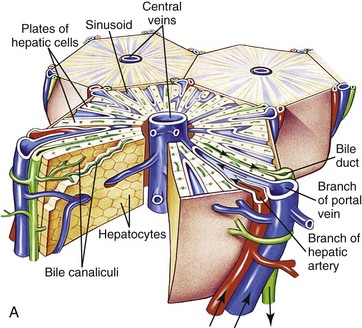
· Microscopic anatomy of the liver (Wikimedia Commons)

· Structure and function of blood vessels (capillary types) (OpenStax Anatomy and Physiology 2e)
Where it is found
Related terms
- Portal triad
- Central vein of the liver
- Interlobular veins of the liver
- Hepatic veins
- Portal vein
- Liver
- Spleen
References and sources
- Further reading: Capillary (sinusoidal capillaries), Wikipedia.
- Further reading: Liver sinusoid, Wikipedia.
- Federative International Programme for Anatomical Terminology (FIPAT). Terminologia Anatomica, 2nd ed. (TA2), 2019.