Atlanto-occipital joint (Latin: articulatio atlantooccipitalis)
What is the atlanto-occipital joint?
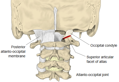
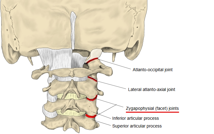
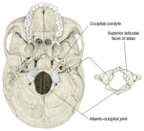
Atlanto-occipital joint: occipital bone (occipital condyle) ⇔ atlas (superior articular facet)
- Each joint is enclosed in a loose joint capsule.
- Taken together, the joints of the two sides act on the head as a single ellipsoid joint with two axes: tilting the head to the side (lateral flexion) about a front-to-back (sagittal) axis, and nodding the head forwards and backwards (flexion and extension) about a side-to-side (transverse) axis.
- The joint is surrounded in front and behind by the anterior and posterior atlanto-occipital membranes, which work like ligaments.
Bones
Ligaments
The two membranes below surround the joint and work like ligaments.
Illustrations
More about the joint
The atlanto-occipital joint lies between the superior articular facets on the lateral masses of the atlas and the occipital condyles, which sit on either side of the foramen magnum of the occipital bone. The facets of the atlas are concave and form the sockets; the condyles are convex and form the heads. The long axes of the right and left joint surfaces, if extended, meet in front of the joint. The facets of the atlas face upwards and medially and have raised outer rims, while the condyles face downwards and laterally, so that the surfaces of both sides fit on one ellipsoid. For this reason the two joints together act as a single ellipsoid joint with two axes: nodding (flexion and extension) about a transverse axis and tilting to the side (lateral flexion) about a sagittal axis.
The joint is strengthened by two membranes:
- Anterior atlanto-occipital membrane: stretches from the anterior arch of the atlas to the anterior margin of the foramen magnum. It contains elastic fibres.
- Posterior atlanto-occipital membrane: stretches from the posterior arch of the atlas to the posterior margin of the foramen magnum and is rich in elastic fibres. At each side it leaves an opening through which the vertebral artery and the first cervical (suboccipital) nerve pass. It is regarded as the upward continuation of the ligamenta flava.