Latin: musculus biceps femoris
Overview
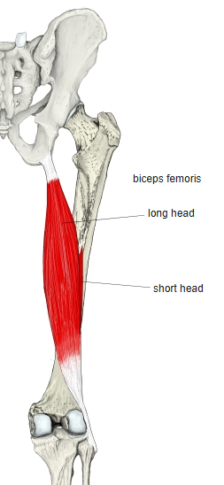
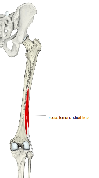
- One of the hamstrings, with a two-joint long head and a one-joint short head.
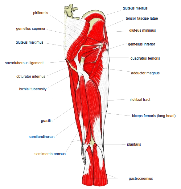
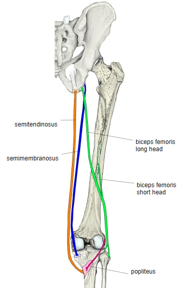
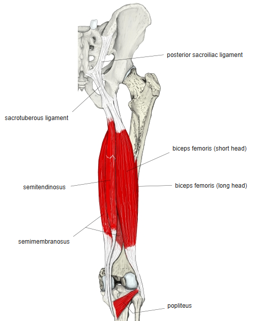
- The long head arises together with the semitendinosus by a common tendon from the ischial tuberosity.
- The two heads unite and insert on the head of the fibula; a bursa lies between the tendon and the fibular collateral ligament of the knee.
- The only lateral rotator of the flexed knee; its rotating action matches that of all the medial rotators combined.
- Continuous in places with:
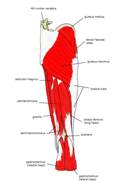
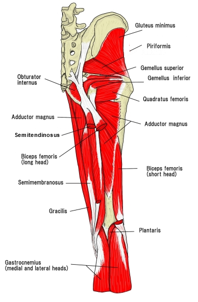
- Long head: gastrocnemius, semitendinosus, fibularis longus
- Short head: vastus intermedius, vastus lateralis, gluteus maximus, adductor magnus, gastrocnemius, popliteus
Position in the classification
Lower Limb › Thigh › Flexor group
- Biceps femoris
- Semitendinosus
- Semimembranosus
Illustrations
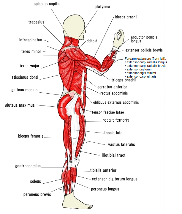
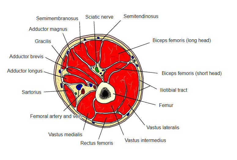
Origin / insertion
- Origin:
- Long head: posterior surface of the ischial tuberosity and the lower part of the sacrotuberous ligament
- Short head: lateral lip of the linea aspera, the lateral supracondylar line and the lateral intermuscular septum
- Insertion: head of the fibula and the crural fascia; a small slip of the tendon reaches the lateral condyle of the tibia
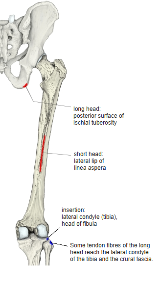
Action
- Flexes the knee and, with the knee flexed, rotates the leg laterally
- Long head: extends the hip
Innervation
- Two nerves:
- Long head: tibial nerve (L5, S1; some sources include S2)
- Short head: common fibular nerve (L4–S1)
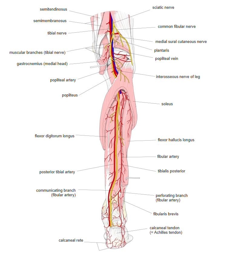
Blood supply
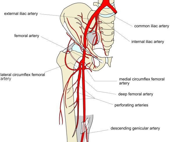
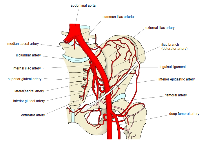
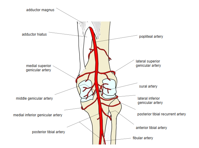
- Perforating arteries of the deep femoral artery, inferior gluteal artery, popliteal artery
Related terms
- Semitendinosus
- Semimembranosus
- Hamstrings
- Ischial tuberosity
- Linea aspera
- Fibula
- Tibial nerve
- Common fibular nerve
References and sources
- Further reading: Biceps femoris muscle, Wikipedia.