Latin: diaphragma
- Overview
- Illustrations
- Origin
- Insertion
- Action
- Innervation
- Blood supply
- Crura and openings
- Structures passing through
Overview
- A dome-shaped sheet of muscle forming the boundary between the thoracic and abdominal cavities.
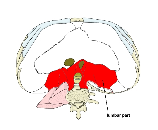
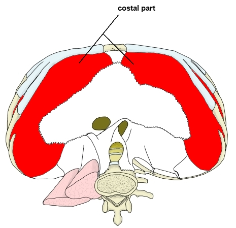
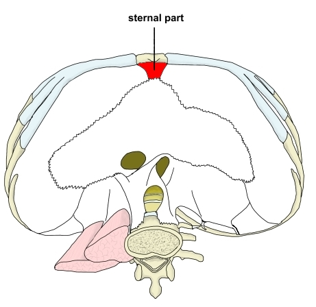
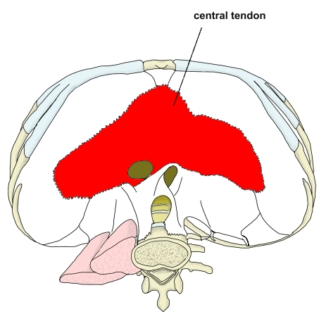
- Three muscular parts – lumbar part (right and left crura), costal part and sternal part – and the central tendon.
- The three muscular parts all insert on the trefoil-shaped central tendon.
- The main muscle of inspiration.
Position in the classification
Trunk › Thorax › Diaphragm
- Diaphragm
Illustrations
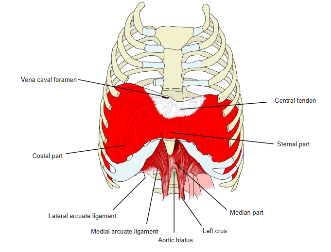
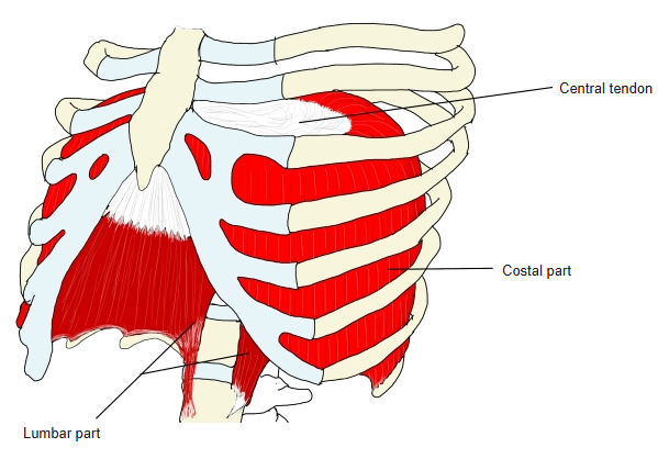
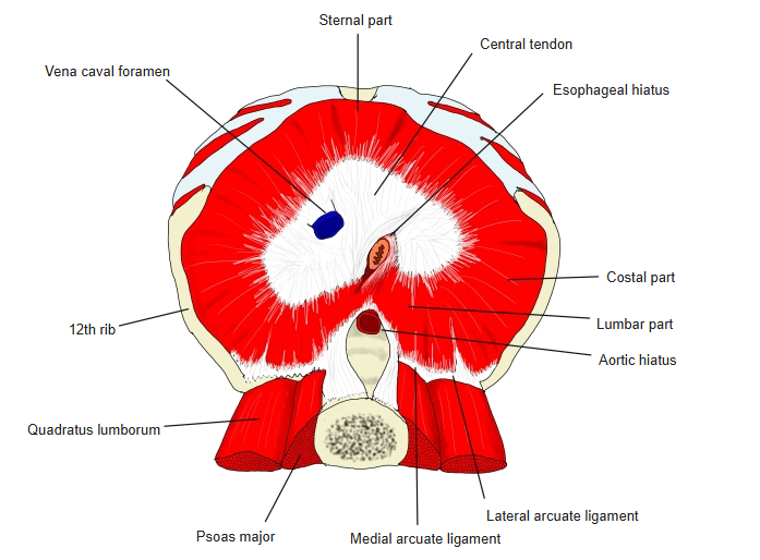
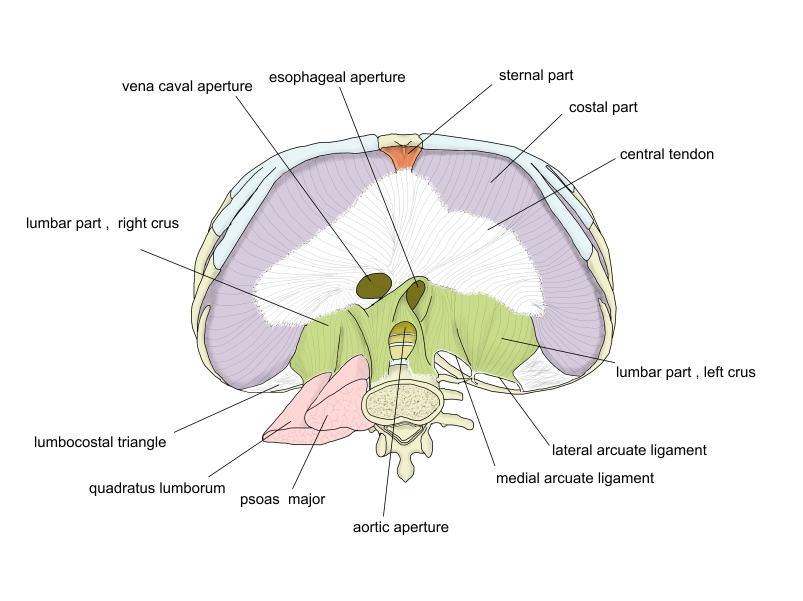
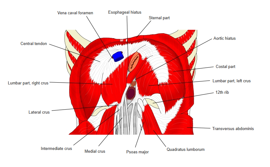
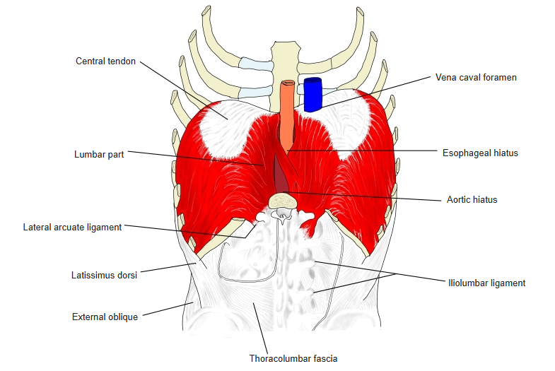
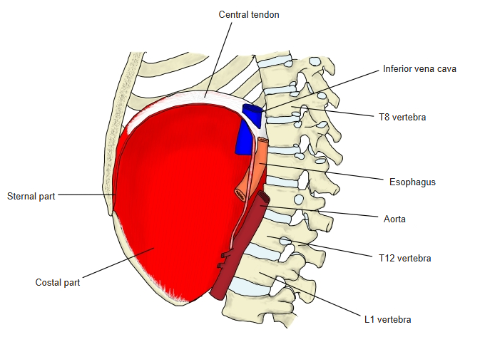
Origin
- Lumbar part
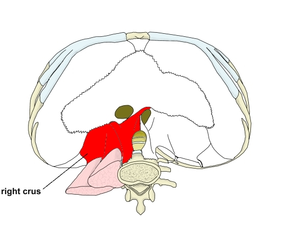
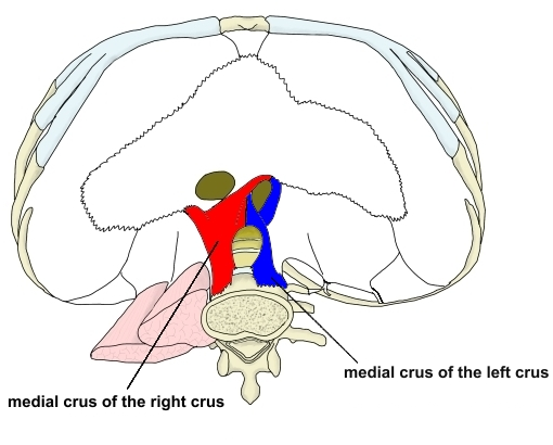
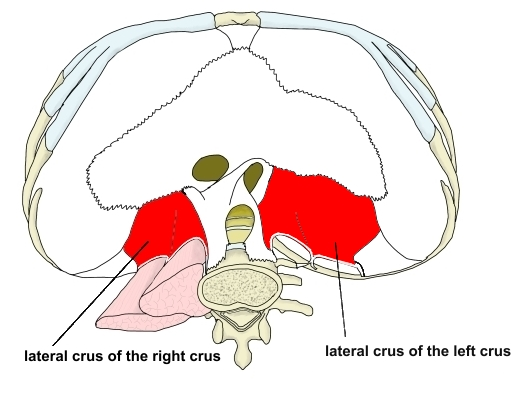
- Right crus – medial crus: bodies of L1–L3; lateral crus: medial and lateral arcuate ligaments
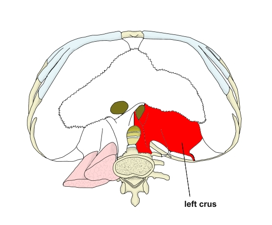
- Left crus – medial crus: bodies of L1–L2; lateral crus: medial and lateral arcuate ligaments
- Costal part: inner surfaces of the 7th–12th costal cartilages and the adjacent parts of the ribs (costal arch)
- Sternal part: posterior surface of the xiphoid process and posterior layer of the rectus sheath
Insertion
- Central tendon – common to all three parts
Action
- Contracts and flattens, enlarging the thoracic cavity for inspiration
Innervation
- Motor: phrenic nerve (C3–C5; chiefly C4)
- Sensory: phrenic and intercostal nerves
- Autonomic: sympathetic (via the cervical ganglia) and parasympathetic (vagus nerve) fibres; regulate blood flow
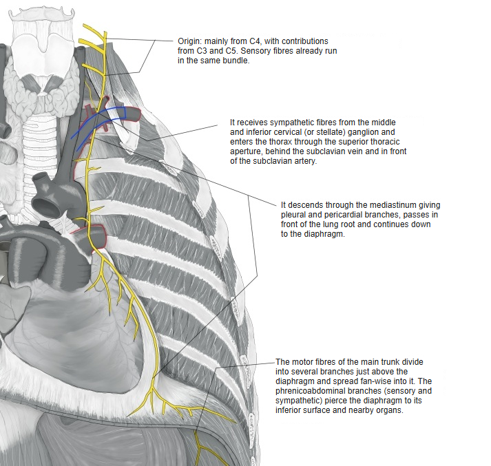
Blood supply
- Arteries
- Inferior phrenic arteries (from the abdominal aorta) – the main supply, reaching the diaphragm from below
- Superior phrenic arteries (from the thoracic aorta)
- Pericardiacophrenic and musculophrenic arteries (from the internal thoracic artery)
- Lower intercostal arteries – along the costal margin
- Veins: run with the arteries; drain to the inferior vena cava, the azygos system and the internal thoracic veins (the left inferior phrenic vein often drains into the left suprarenal vein)
Compiled from standard anatomy references.
Crura, arcuate ligaments and openings
- Right / left crus: the parts of the lumbar part to the right and left of the vertebral column
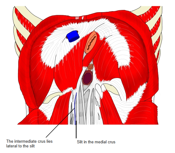
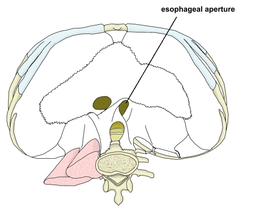
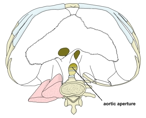
- Medial crus: arises from the lumbar vertebral bodies; the medial margins of the two crura meet in front of the aorta, forming an arch (the median arcuate ligament) over the aortic hiatus; the esophageal hiatus lies above, in front of and slightly to the left of it, and is formed mainly by fibres of the right crus
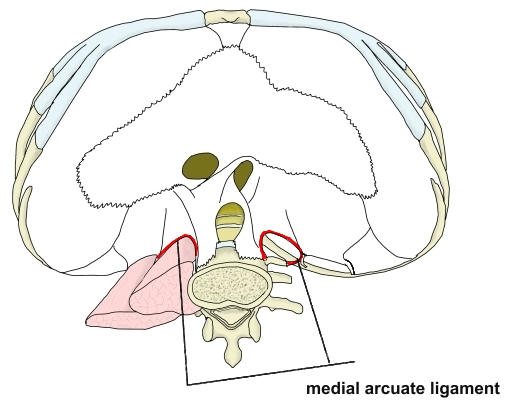
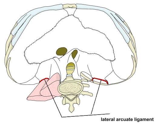
- Lateral crus: arises from the medial and lateral arcuate ligaments
- Intermediate crus: the part lateral to a slit in the medial crus
- Medial arcuate ligament: between the body and costal process of L1 (or L2), over the psoas major
- Lateral arcuate ligament: between the costal process of L1 (or L2) and the tip of the 12th rib, over the quadratus lumborum
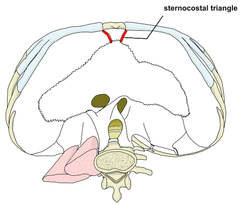
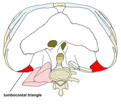
- Sternocostal / lumbocostal triangles: gaps without muscle fibres between the costal and sternal parts, and between the lateral arcuate ligament and the costal part
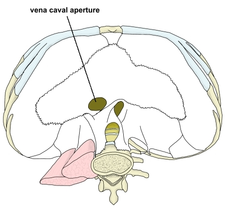
- Openings: vena caval foramen (in the central tendon, about the level of T8), esophageal hiatus (about T10) and aortic hiatus (about T12)
Structures passing through
| Opening or gap | Structures |
|---|---|
| Vena caval foramen | Inferior vena cava; branches of the right phrenic nerve |
| Esophageal hiatus | Esophagus; vagus nerves |
| Aortic hiatus | Descending aorta; thoracic duct; azygos vein |
| Slit in the medial crus | Greater splanchnic nerve; hemiazygos vein (left); occasionally the azygos vein (right) |
| Intermediate crus | Lesser splanchnic nerve |
| Between the medial and lateral crus (behind the medial arcuate ligament) | Sympathetic trunk |
| Sternocostal triangle | Superior epigastric artery and vein; lymphatic vessels |
Related terms
- Central tendon
- Right crus
- Left crus
- Medial arcuate ligament
- Lateral arcuate ligament
- Vena caval foramen
- Esophageal hiatus
- Aortic hiatus
- Phrenic nerve
- Respiratory muscles
- External intercostal muscles
- Psoas major
- Quadratus lumborum
References and sources
- Further reading: Thoracic diaphragm, Wikipedia.
- Blood supply: The Diaphragm, TeachMeAnatomy; and Wikipedia (above).