Latin: musculus extensor hallucis brevis
Overview
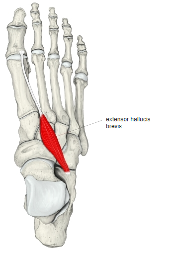
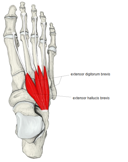
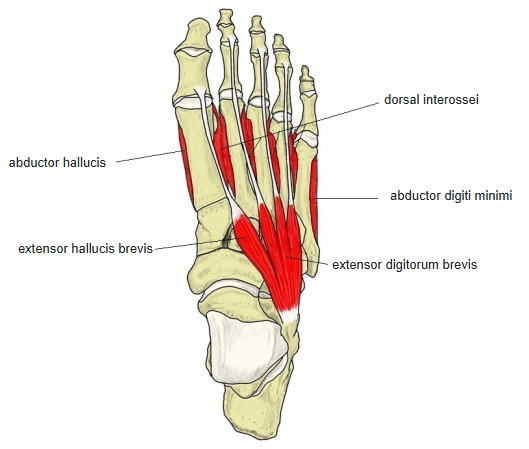
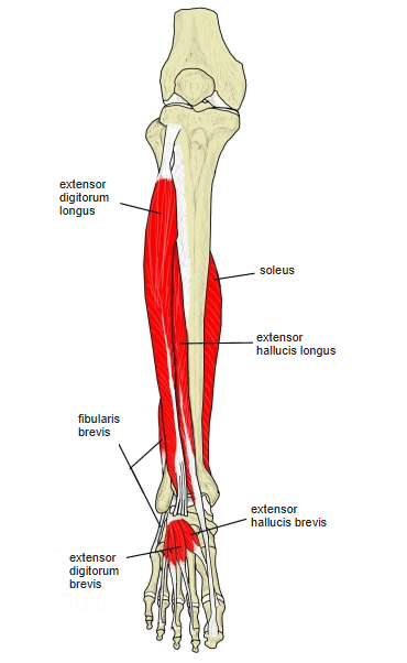
- It is essentially the medial part of the extensor digitorum brevis; it is separated from that muscle and shares its origin on the calcaneus.
- Some anatomists regard the two as a single muscle.
Position in the classification
Lower Limb › Foot › Dorsum of foot
- Extensor digitorum brevis
- Extensor hallucis brevis
Illustrations
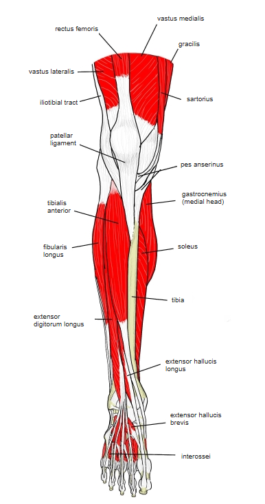
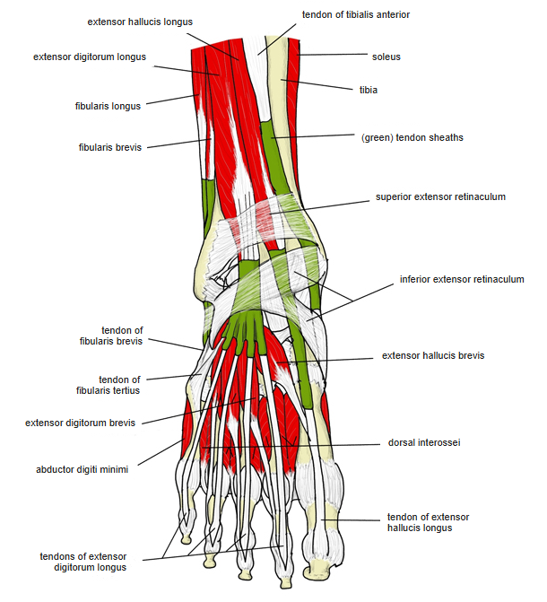
Origin / insertion
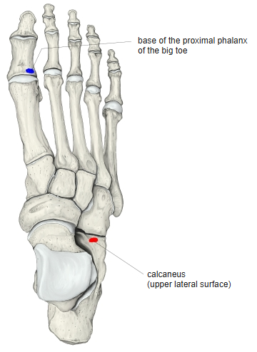
- Origin: upper and lateral surfaces of the calcaneus (with the extensor digitorum brevis); some sources add the talocalcaneal ligament and the stem of the inferior extensor retinaculum
- Insertion: dorsal surface of the base of the proximal phalanx of the great toe (some sources add the dorsal aponeurosis of the great toe)
Action
- Extends the great toe at the metatarsophalangeal joint
Innervation
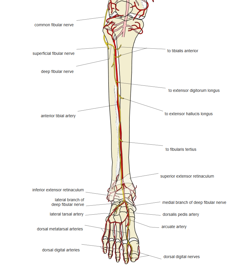
- Deep fibular nerve (L5, S1; some sources L4–S1)
Blood supply
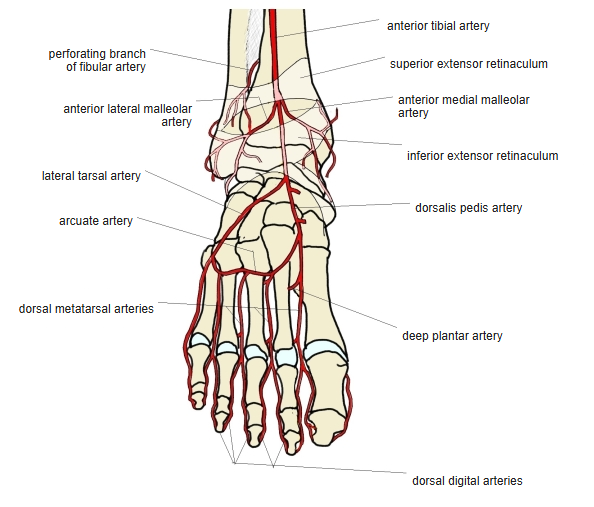
- Branches of the dorsalis pedis artery
Related terms
- Extensor digitorum brevis
- Extensor hallucis longus
- Calcaneus
- Deep fibular nerve
- Dorsalis pedis artery
References and sources
- Further reading: Extensor hallucis brevis muscle, Wikipedia.