Latin: musculus flexor digitorum superficialis
Overview
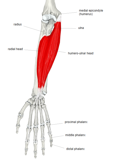
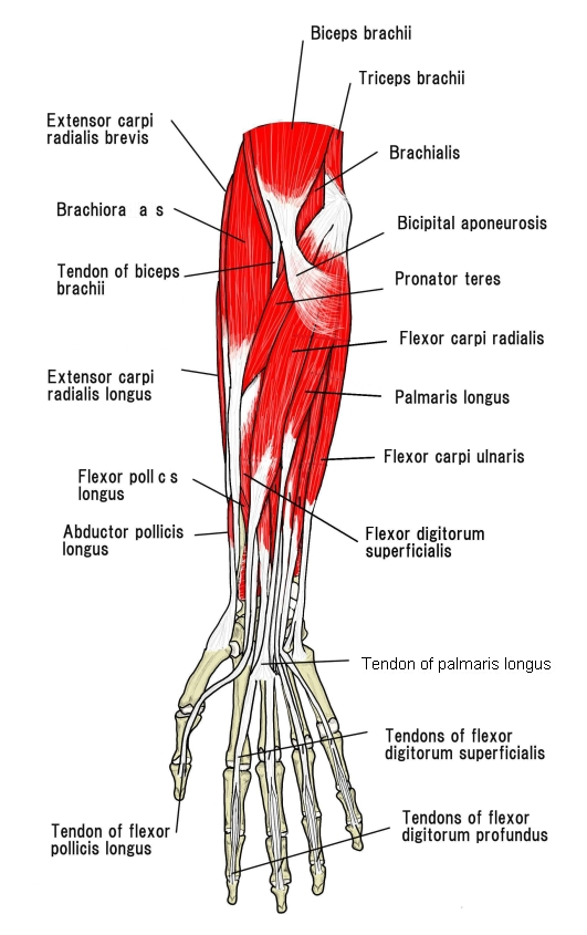
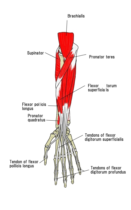
- One of the 8 flexor muscles of the forearm, in the second (middle) layer; most of its belly is covered by the first-layer muscles.
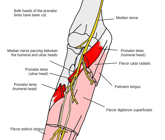
- Two heads, humero-ulnar and radial; the median nerve and ulnar artery pass between them.
- The broad belly divides into four tendons: those to the middle and ring fingers lie superficially, those to the index and little fingers deeper. All pass through the carpal tunnel.
- Over the proximal phalanx each tendon splits to let the deep flexor tendon through, and part of the fibres cross (tendinous chiasm).
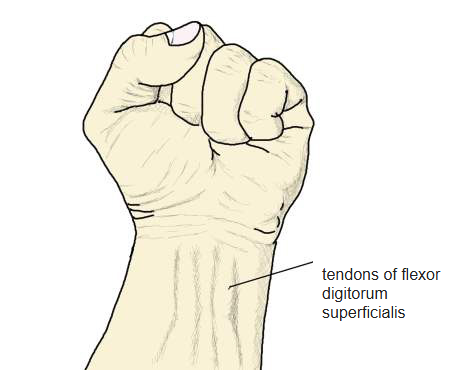
- Palpation: make a fist and flex the wrist hard; the tendons of the flexor carpi radialis, palmaris longus and this muscle stand out.
- Continuous in places with the pronator teres, biceps brachii, flexor carpi ulnaris, flexor carpi radialis and palmaris longus.
Position in the classification
Upper Limb › Forearm › Flexor group › Second and third layers
- Flexor digitorum superficialis
- Flexor digitorum profundus
- Flexor pollicis longus
Illustrations
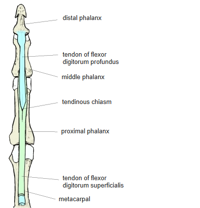
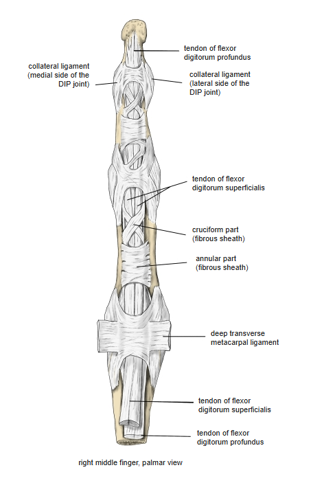
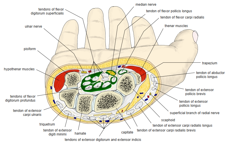
Origin / insertion
- Origin:
- Humero-ulnar head: medial epicondyle of the humerus (common flexor tendon), the ulnar collateral ligament of the elbow and the medial side of the coronoid process of the ulna (some sources: tuberosity of the ulna)
- Radial head: oblique line (anterior border) of the radius, from the radial tuberosity to the insertion of the pronator teres
- Insertion: sides of the middle phalanges of the 2nd to 5th fingers (each tendon splits into two slips)
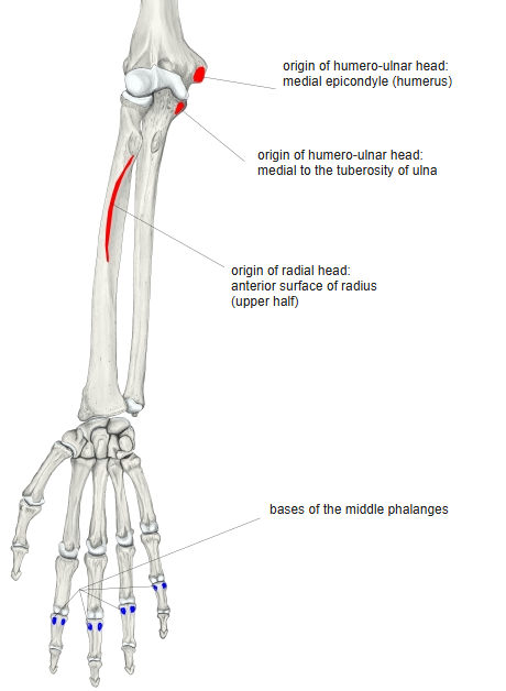
Action
- Flexes the proximal interphalangeal (PIP) joints of the 2nd to 5th fingers
- Also helps to flex the metacarpophalangeal joints and the wrist
Innervation
- Median nerve (C7–T1)
Blood supply
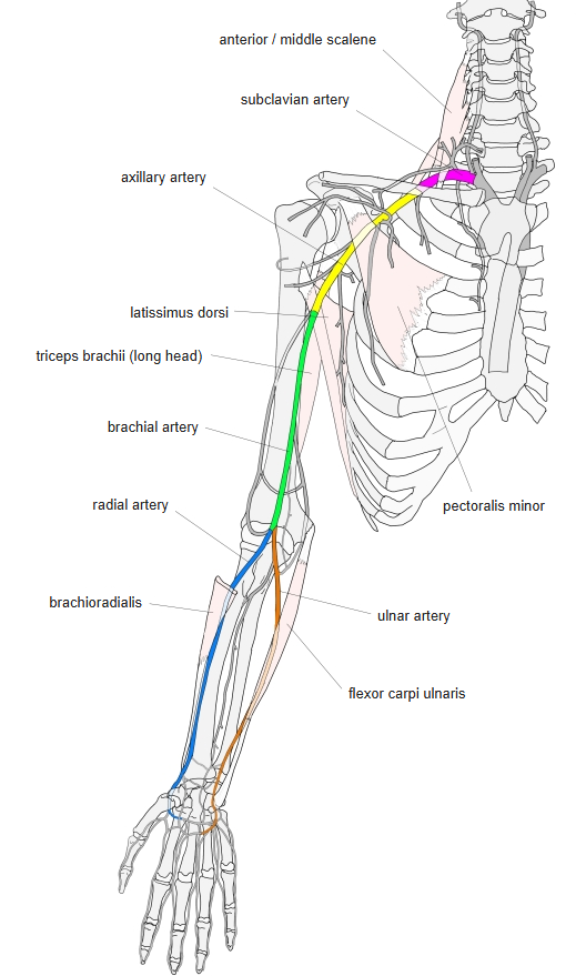
- Ulnar artery, radial artery
Anatomical variations
- May split into four separate muscles; the radial head may be small or absent
- The tendon to the little finger is often missing (on one or both sides)
- The belly to the index finger is often separate
- A tendon may join the palmar aponeurosis; slips may come from the flexor digitorum profundus
Related terms
- Flexor digitorum profundus
- Pronator teres
- Palmaris longus
- Flexor carpi radialis
- Medial epicondyle
- Carpal tunnel
- Median nerve
- Ulnar artery
References and sources
- Further reading: Flexor digitorum superficialis muscle, Wikipedia.