Latin: musculus gastrocnemius
Overview
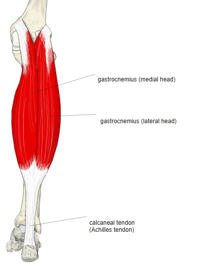
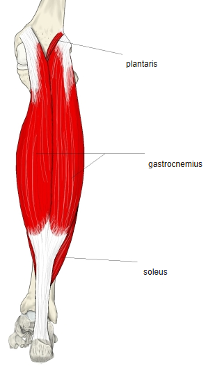
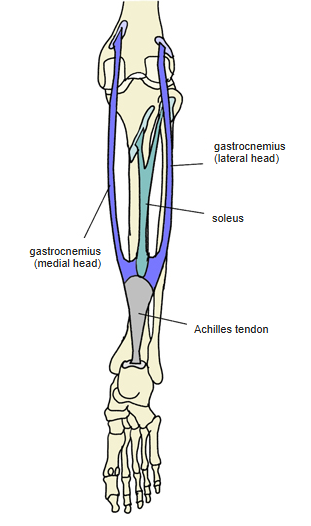
- The most superficial muscle of the calf; together with the soleus it forms the triceps surae.
- Gastrocnemius compared with the soleus:
- Gastrocnemius: mostly fast fibres; crosses two joints (knee and ankle)
- Soleus: mostly slow fibres; crosses one joint (ankle)
- Some sources also count the subtalar joint, making it a three-joint muscle.
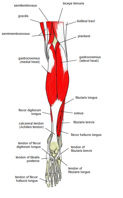
- Its origins lie about 5 cm above the lower end of the femur, the medial head slightly higher; many fibres arise directly from the femur and are covered by the semimembranosus and biceps femoris.
- A sesamoid bone or cartilage, the fabella, occurs in it in about 35% (usually on both sides); the lateral head may be absent or rudimentary.
- A small bursa lies between the back of the calcaneus and its tendon.
- Continuous in places with the biceps femoris, adductor magnus, flexor digitorum longus, soleus and plantaris.
Position in the classification
Lower Limb › Leg › Flexor group › Triceps surae
- Gastrocnemius
- Soleus
Illustrations
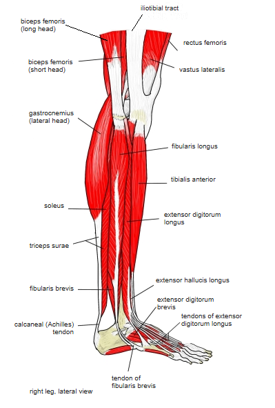
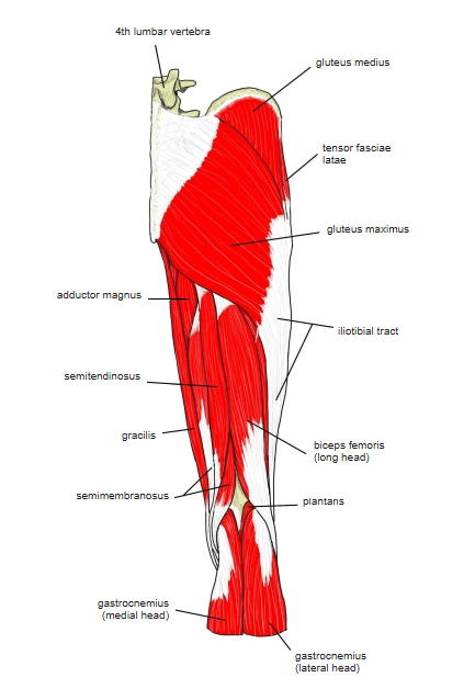
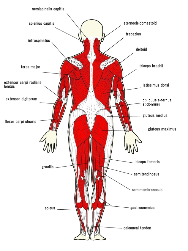
Origin / insertion
- Origin:
- Medial head: upper posterior part of the medial condyle of the femur and the adjacent femur
- Lateral head: lateral side of the lateral condyle of the femur and the posterior surface of the femur just above it; both heads also arise from the knee joint capsule
- Insertion: calcaneal tuberosity, by the calcaneal (Achilles) tendon shared with the soleus
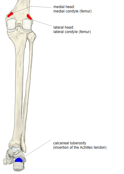
Action
- Plantar flexes the foot, raises the heel and flexes the knee
- With the foot fixed, draws the leg and thigh backwards
Innervation
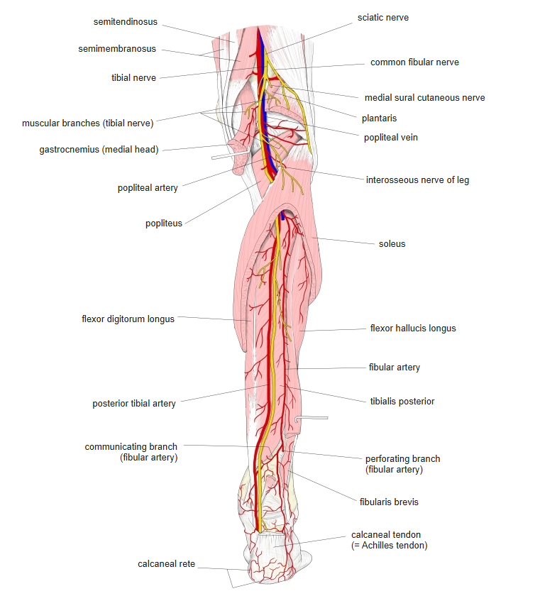
- Tibial nerve (S1, S2)
Blood supply
- Sural arteries (branches of the popliteal artery)
Related terms
- Soleus
- Plantaris
- Triceps surae
- Calcaneal tendon (Achilles tendon)
- Fabella
- Femur
- Tibial nerve
References and sources
- Further reading: Gastrocnemius muscle, Wikipedia.