Latin: musculus levator palpebrae superioris
Overview
- Listed here as one of the 7 extraocular muscles, although it does not move the eyeball.
- Some sources exclude it from the extraocular muscles and list only the four recti and two obliques, which move the eyeball.
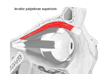
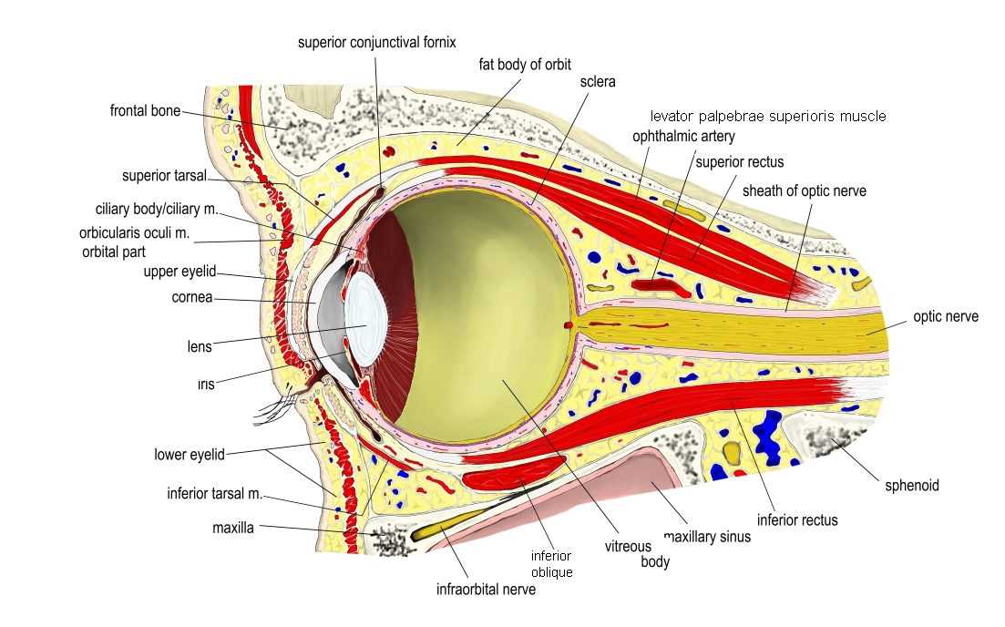
- Lies in the orbit just under its roof, above the superior rectus; the superior oblique lies at the upper medial side of the orbit.
- Widens and thins toward the front into the levator aponeurosis.
- Smooth muscle fibres in the middle layer of its aponeurosis form the superior tarsal muscle (Müller's muscle), which inserts into the upper margin of the superior tarsus.
Position in the classification
Head › Extraocular muscles
- Superior rectus
- Inferior rectus
- Medial rectus
- Lateral rectus
- Superior oblique
- Inferior oblique
- Levator palpebrae superioris
Illustrations
Origin / insertion
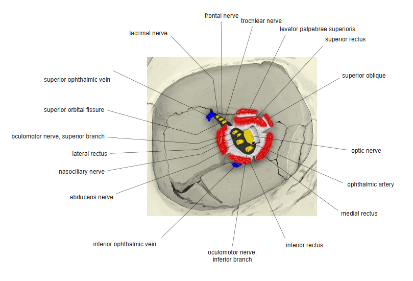
- Origin: under surface of the lesser wing of the sphenoid, above and in front of the optic canal
- Runs forward just below the roof of the orbit.
- Insertion: through its aponeurosis to the skin of the upper eyelid, the upper margin of the superior tarsus and the superior fornix of the conjunctiva
Action
- Raises the upper eyelid; it is the direct antagonist of the orbicularis oculi
Innervation
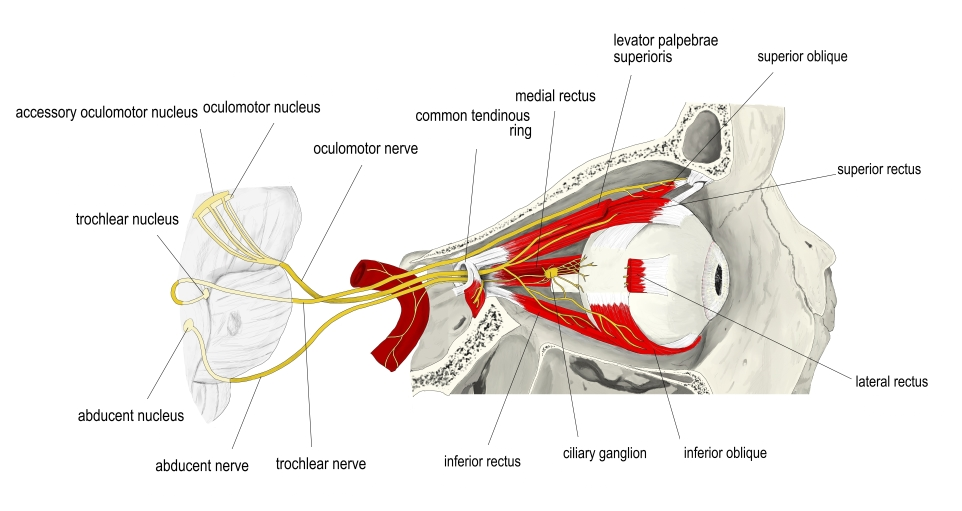
- Oculomotor nerve (cranial nerve III), superior branch
- The superior tarsal muscle (smooth muscle) is supplied by sympathetic fibres from the superior cervical ganglion.
Blood supply
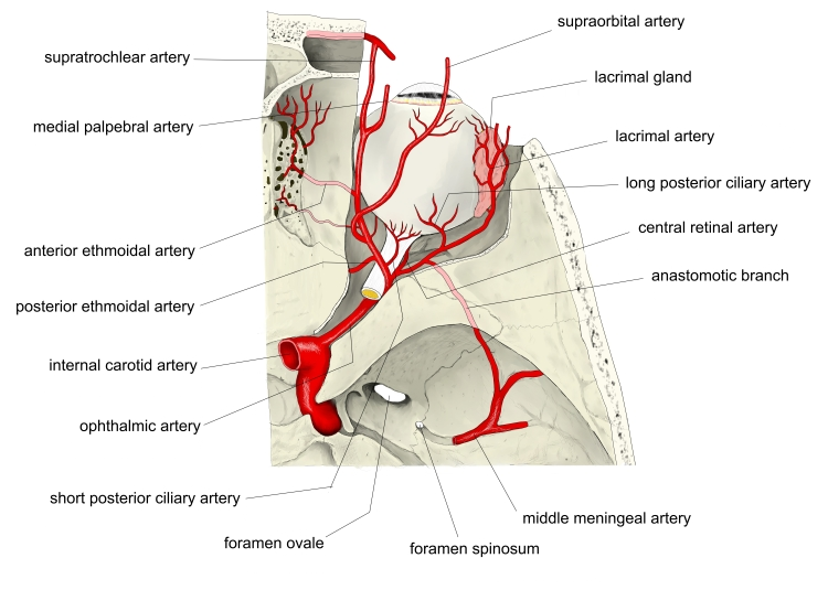
- Arteries: supraorbital artery and muscular branches of the ophthalmic artery
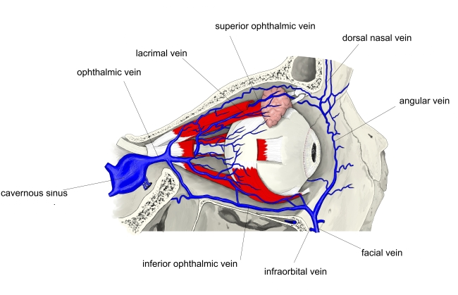
- Vein: superior ophthalmic vein (drains to the cavernous sinus)
Related terms
- Extraocular muscles
- Superior rectus
- Superior oblique
- Superior tarsal muscle
- Tarsal plate
- Lesser wing of sphenoid
- Oculomotor nerve
- Ophthalmic artery
- Superior ophthalmic vein
References and sources
- Further reading: Levator palpebrae superioris muscle, Wikipedia.
- Gray H. Anatomy of the Human Body, 20th ed. (1918), “1c. 3. The Accessory Organs of the Eye”, Bartleby.com.