Latin: musculi lumbricales manus
Overview
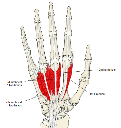
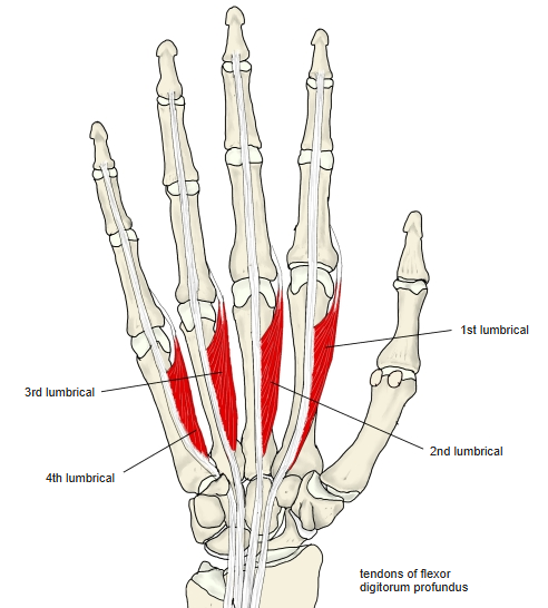
- Four small, worm-like muscles that attach to tendons, not bone.
- Heads:
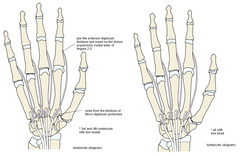
- 1st and 2nd: one head each
- 3rd and 4th: usually two heads
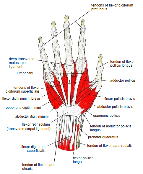
- They run on the palmar side of the deep transverse metacarpal ligament, i.e. in front of the flexion axis of the MP joints.
- They contribute to precise finger movements such as pinching; they cannot be palpated individually.
Position in the classification
Upper Limb › Hand › Metacarpal muscles
- Lumbricals (hand)
- Dorsal interossei (hand)
- Palmar interossei
Illustrations
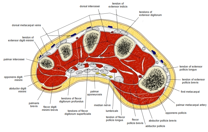
Origin / insertion
- Origin: tendons of the flexor digitorum profundus
- One head: from the radial side of a tendon (1st and 2nd: tendons of the index and middle fingers)
- Two heads: from the facing sides of two adjacent tendons (3rd: middle and ring fingers; 4th: ring and little fingers)
- Insertion: radial side of the dorsal (extensor) expansions of the 2nd–5th fingers, at the level of the proximal phalanges
Action
- Flex the metacarpophalangeal (MP) joints of the 2nd–5th fingers
- Extend the interphalangeal (PIP, DIP) joints
Innervation
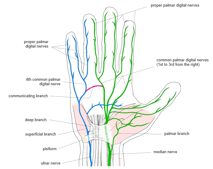
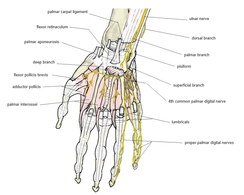
- 1st and 2nd: median nerve; 3rd and 4th: deep branch of the ulnar nerve; segments C8, T1
- This 2:2 pattern is found in about 60%; the 1:3 and 3:1 patterns in about 20% each
Blood supply
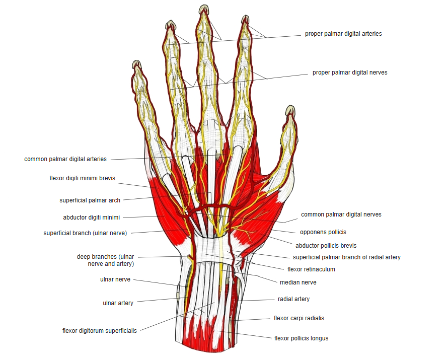
- Superficial palmar arch
Related terms
- Flexor digitorum profundus
- Dorsal interossei (hand)
- Palmar interossei
- Lumbricals (foot)
- Median nerve
- Ulnar nerve
References and sources
- Further reading: Lumbricals of the hand, Wikipedia.
- Gray H. Anatomy of the Human Body, 20th ed. (1918), “1F. The Muscles and Fasciæ of the Hand”, Bartleby.com.