Latin: musculus triceps brachii
Overview
- One of the 2 extensor muscles of the arm (with the anconeus); the large muscle of the back of the arm.
- Has three heads: long, lateral and medial. The medial head is mostly covered by the other two.
- The antagonist of the biceps brachii and brachialis.
- The long head passes between the teres minor (behind) and teres major (in front).
- Deep fibres attach to the elbow-joint capsule (sometimes called the articularis cubiti).
- Continuous in places with the brachialis, deltoid, brachioradialis, coracobrachialis, teres minor and extensor carpi radialis longus.
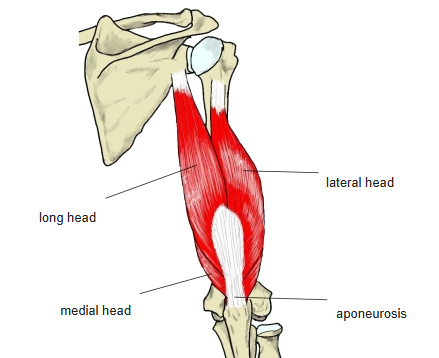
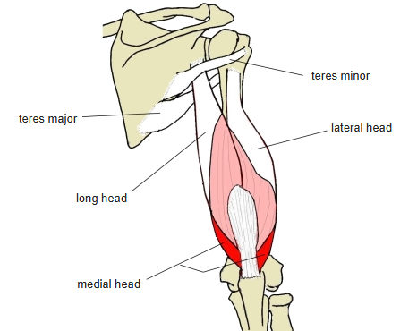
Position in the classification
Upper Limb › Arm › Extensor group
- Triceps brachii
- Anconeus
Illustrations
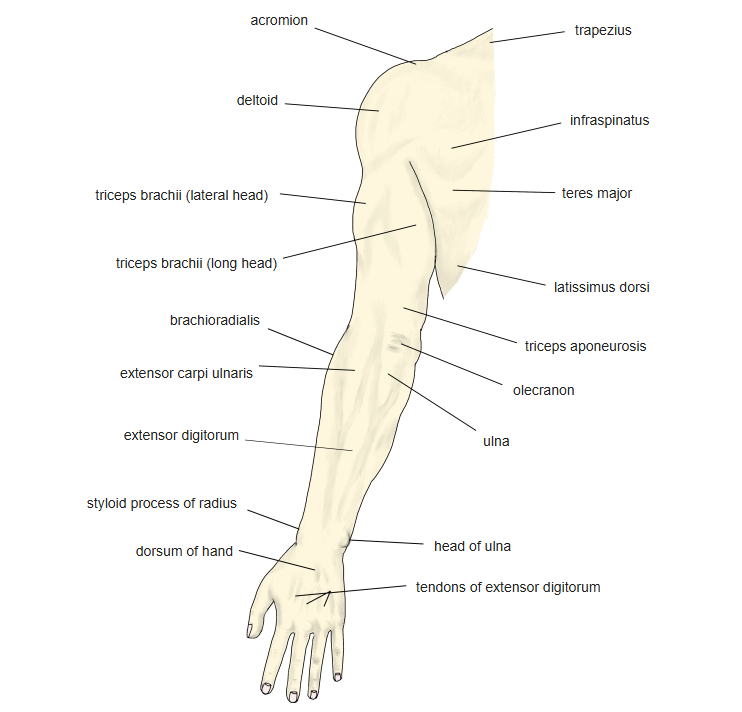
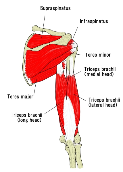
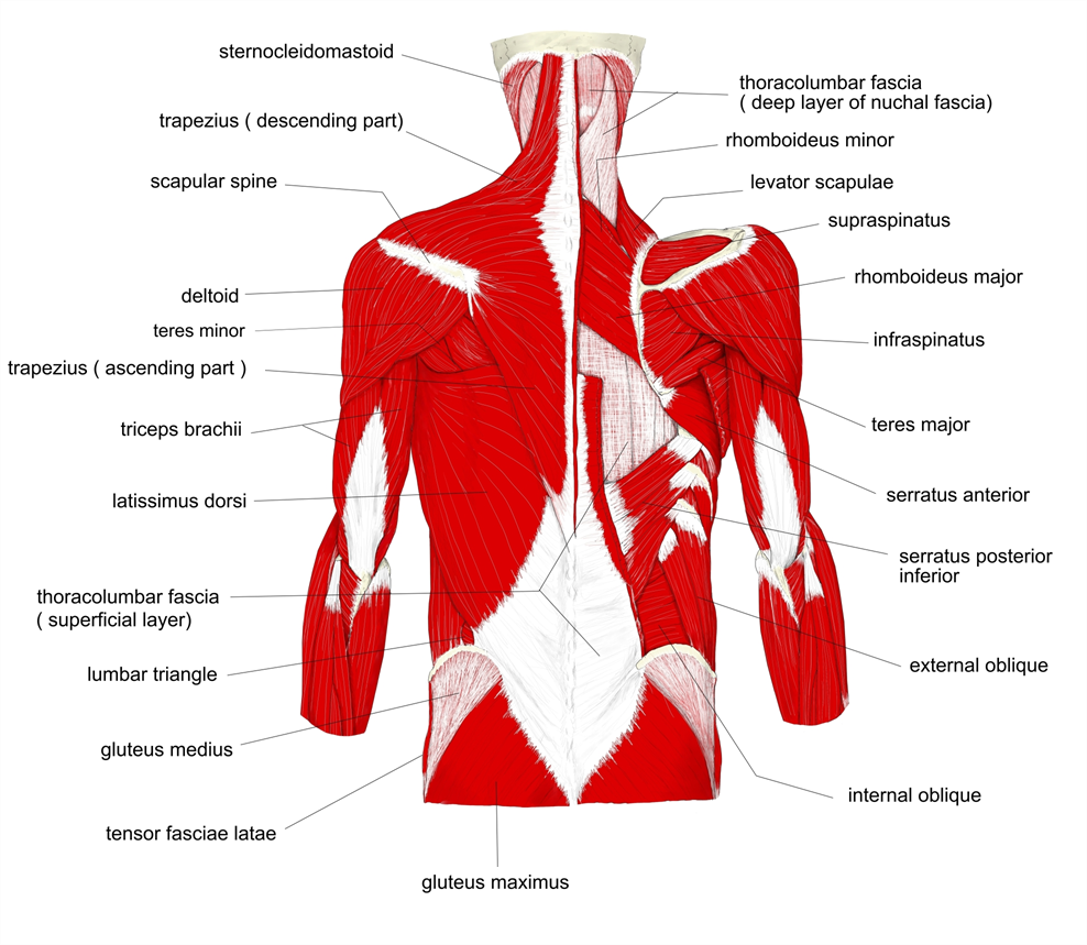
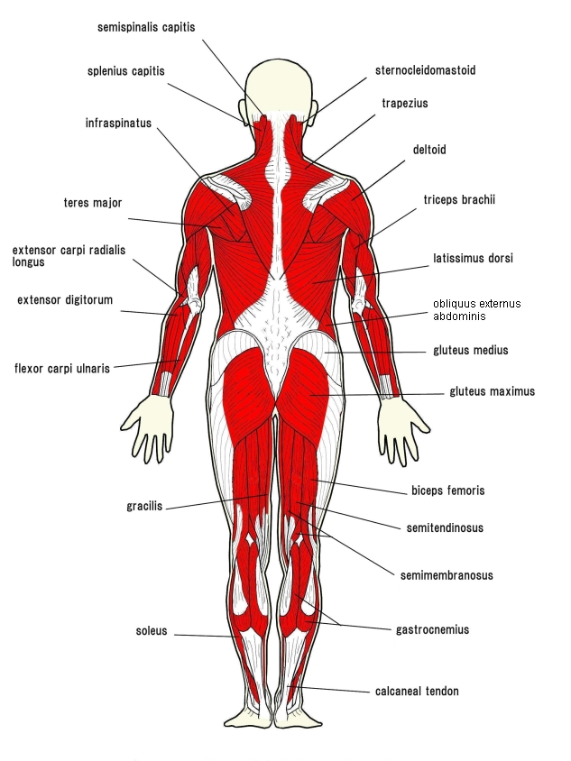
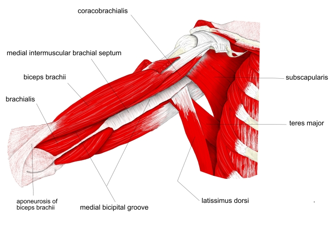
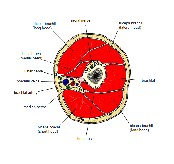
Origin / insertion
- Origin:
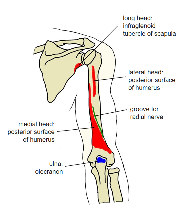
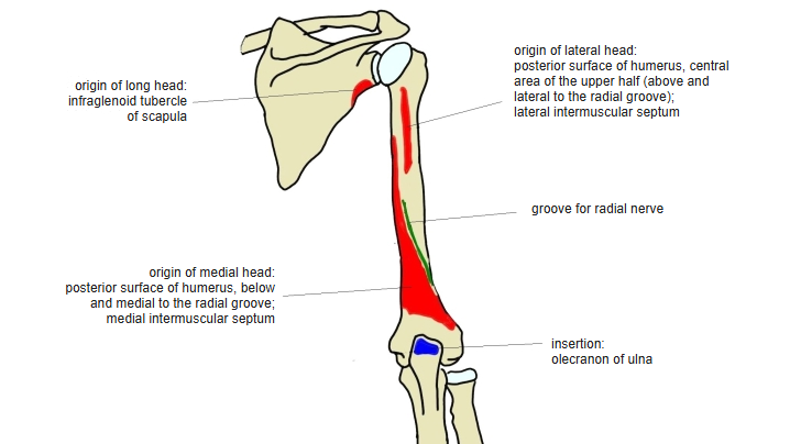
- Long head: infraglenoid tubercle of the scapula (and the adjoining lateral border)
- Lateral head: posterior surface of the humerus, central area of the upper half (above and lateral to the groove for the radial nerve), and the lateral intermuscular septum
- Medial head: posterior surface of the humerus below and medial to the groove for the radial nerve, and the medial (and lateral) intermuscular septum
- Insertion: olecranon of the ulna, by a broad tendinous sheet; lateral fibres continue into the antebrachial fascia
Action
- Extends the elbow
- Light extension: mainly the medial head; forceful extension: the long and lateral heads join in
- The medial and lateral heads extend the elbow to a right angle; the long head completes full extension
- The long head also adducts (and slightly extends) the arm at the shoulder
Innervation
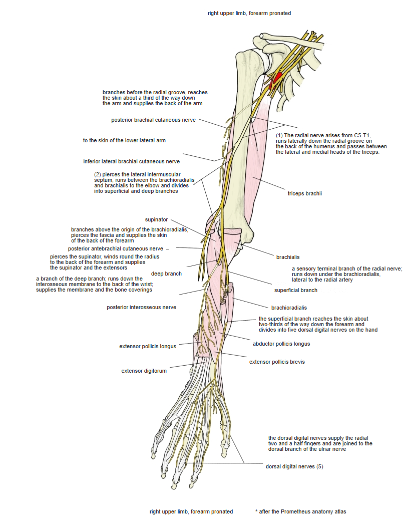
- Radial nerve (C6–C8)
Blood supply
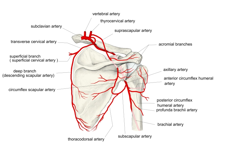
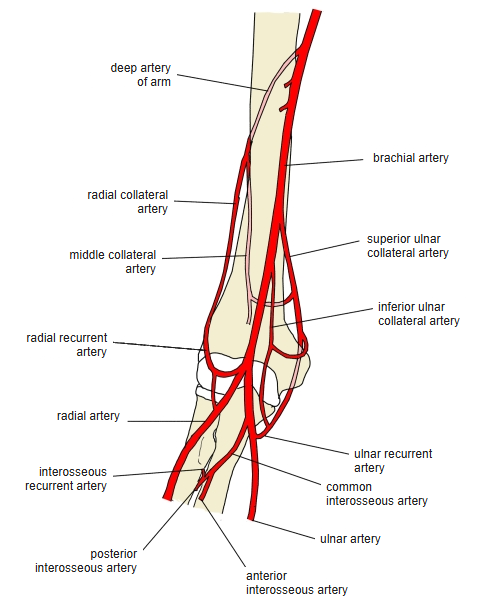
- Posterior circumflex humeral artery, deep artery of the arm; probably also the superior and inferior ulnar collateral arteries
Anatomical variations
- May have a fourth head, from the lateral border of the scapula, the coracoid process, the shoulder-joint capsule or the humerus
- May be joined to the subscapularis, latissimus dorsi or teres major; the origin tendon of the long head is almost always linked to the latissimus dorsi tendon (Krause)
Related terms
- Biceps brachii
- Brachialis
- Anconeus
- Teres minor
- Teres major
- Infraglenoid tubercle
- Olecranon
- Radial nerve
- Deep artery of arm
- Posterior circumflex humeral artery
References and sources
- Further reading: Triceps, Wikipedia.