Latin: pupilla
Overview
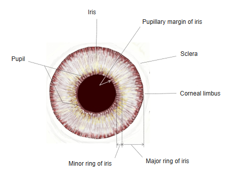
- The round opening in the centre of the iris, lying slightly to the nasal side; it looks black because the inside of the eye absorbs the light.
- Light passes through it to the lens; it also connects the anterior and posterior chambers.
- Its diameter changes from about 1–2 mm in bright light to about 8 mm in the dark.
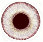
Iris and pupil, anterior view 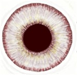
Iris and pupil
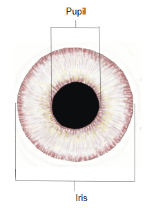
Changes in size
- Light: bright light narrows the pupil (miosis), darkness widens it (mydriasis) – the pupillary light reflex; both pupils react together.
- Near vision: looking at a near object narrows the pupil, together with accommodation and convergence (near response).
- Emotion and drugs: sympathetic activity (fear, excitement) widens it; many drugs narrow or widen it.
- Age: the pupil is smaller in old age.

Size of the pupil
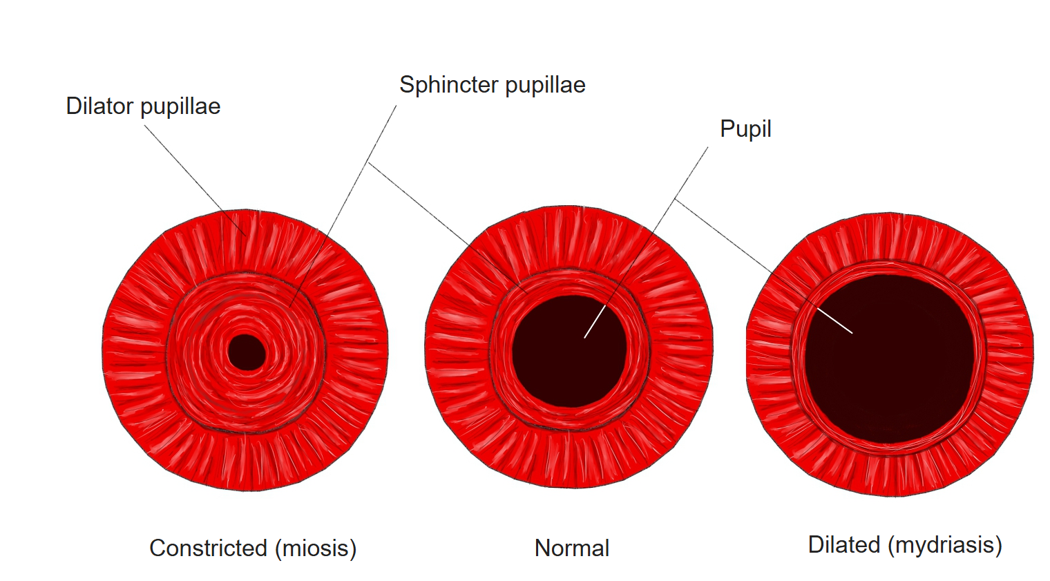
Muscles of the pupil
| Muscle | Action | Nerve supply |
|---|---|---|
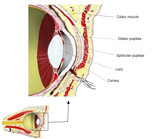
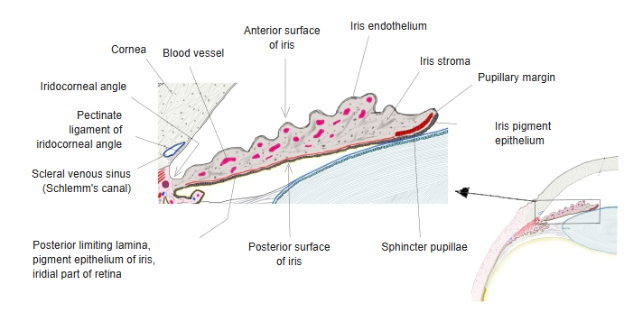
| Sphincter pupillae | Narrows the pupil | Parasympathetic: oculomotor nerve → ciliary ganglion → short ciliary nerves |
| Dilator pupillae | Widens the pupil | Sympathetic: superior cervical ganglion → long ciliary nerves |
- Both muscles of the iris develop from the epithelium (neuroectoderm), not from mesoderm like other smooth muscle.
Intraocular muscles 
Iris, section
References and sources
- Further reading: Pupil, Wikipedia.