Latin: organum spirale
Overview
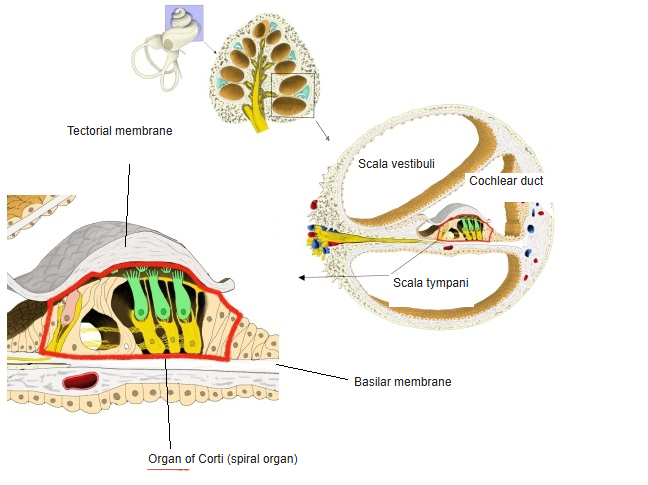
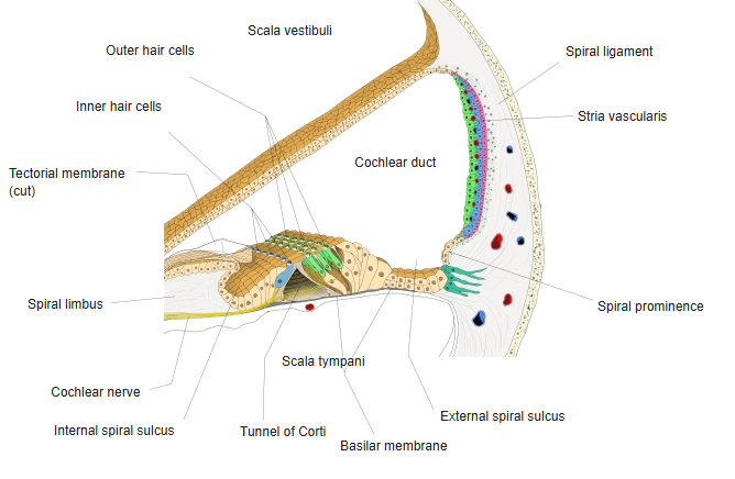
- The organ of hearing: a ridge of sensory and supporting cells on the basilar membrane of the cochlear duct.
- Runs the whole length of the cochlear duct.
- Its hair cells turn the vibration of the basilar membrane into nerve signals.
- The tectorial membrane lies over the hair cells; their stereocilia are bent as the basilar membrane moves against it.
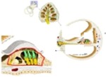
Position of the spiral organ 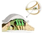
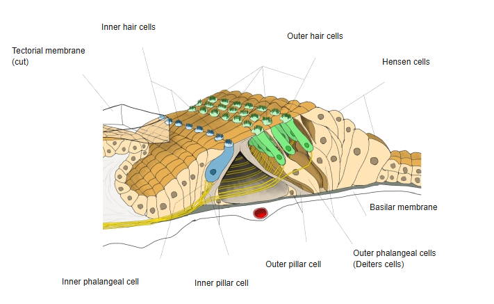
Spiral organ (organ of Corti)
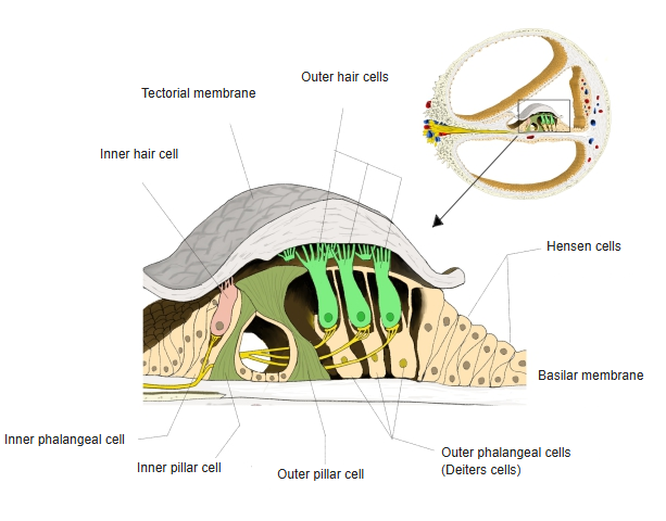
Cells
| Cells | Description |
|---|---|
| Inner hair cells | One row, about 3,500 cells; flask-shaped; the main sensory cells – most fibres of the cochlear nerve end on them |
| Outer hair cells | Three (to four) rows, about 12,000 cells; cylindrical; they change length and amplify the movement of the basilar membrane |
| Inner and outer pillar cells | Stiff supporting cells that enclose the triangular tunnel of Corti |
| Inner and outer phalangeal cells | Supporting cells holding the hair cells; the outer ones are the Deiters cells |
| Hensen cells, Claudius cells | Tall and then cuboidal border cells lateral to the outer hair cells |
- High-frequency sound moves the basilar membrane near the base of the cochlea, low-frequency sound near the apex (tonotopy).
- Signals pass from the hair cells to the bipolar cells of the spiral ganglion and on in the cochlear nerve.
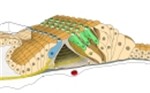
Spiral organ, three-dimensional view 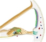
Cochlear duct, cross-section
References and sources
- Further reading: Spiral organ (organ of Corti), Wikipedia.