Latin: utriculus
Overview
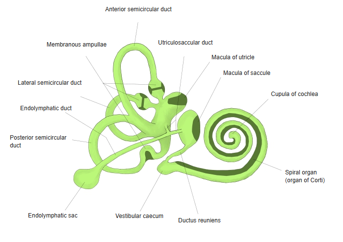
- The larger of the two membranous sacs in the vestibule, lying in the elliptical recess.
- The three semicircular ducts open into it by five openings.
- Its sensory area, the macula of the utricle, lies roughly horizontal on its floor and senses tilt of the head and horizontal linear acceleration.
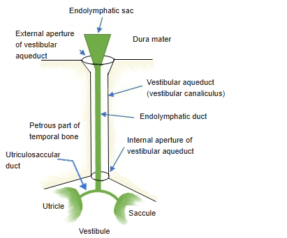
- Connected with the saccule by the utriculosaccular duct, which gives off the endolymphatic duct.
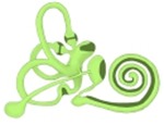
Membranous labyrinth 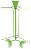
Endolymphatic duct and sac (schematic)
Structure of the macula
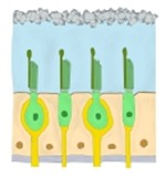
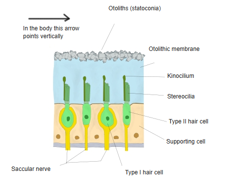
- A thickened area of epithelium with hair cells (type I and type II) and supporting cells.
- Each hair cell carries many stereocilia and one kinocilium, which project into the gelatinous otolithic membrane.
- On the membrane lie calcium carbonate crystals, the otoliths (statoconia); their weight makes the membrane slide when the head tilts, bending the hair bundles.
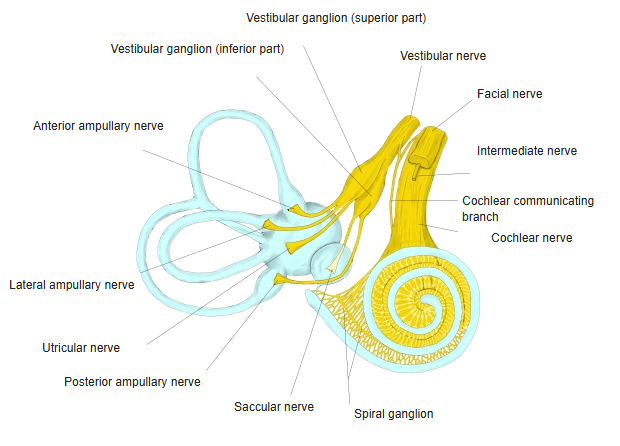
- Fibres from the macula run in the utricular nerve, part of the vestibular nerve.
Macula, structure 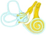
Nerves of the membranous labyrinth
References and sources
- Further reading: Utricle, Wikipedia.