Latin: nervus phrenicus
Overview
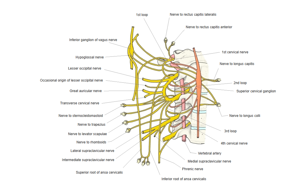
- Arises mainly from C4, with fibres from C3 and C5; the only motor nerve of the diaphragm.
- A mixed nerve: mostly motor, with sensory fibres to the pleura, pericardium and peritoneum and sympathetic fibres added in the neck.
- There is a right and a left phrenic nerve.
Cervical plexus (schematic)
Course
- Descends obliquely on the front of scalenus anterior, from its lateral to its medial border, deep to the prevertebral fascia.
- Low in the neck it receives sympathetic fibres from the middle or inferior cervical (stellate) ganglion.
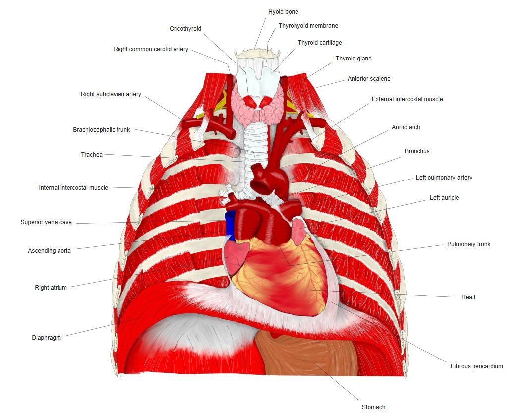
- Enters the thorax in front of the subclavian artery and behind the subclavian vein.
- In the mediastinum it runs between the mediastinal pleura and the fibrous pericardium with the pericardiacophrenic vessels, in front of the root of the lung.
- The left nerve slopes forwards more because it follows the bulge of the heart.
- At the diaphragm it divides; branches spread on its upper surface, and the phrenicoabdominal branches pass through the caval opening (right) or the muscle (left) to its under surface.
Neck and thorax (front view)
Branches and supply
- An accessory phrenic nerve, usually from the nerve to subclavius (C5), may join it.
| Branch | Supply |
|---|---|
| Motor branches | Diaphragm |
| Pleural branches | Mediastinal and diaphragmatic pleura (sensory) |
| Pericardial branch | Fibrous and parietal serous pericardium (sensory) |
| Phrenicoabdominal branches | Peritoneum under the diaphragm, nearby ligaments, inferior vena cava and suprarenal region; join the phrenic plexus |
References and sources
- Further reading: Phrenic nerve, Wikipedia.