Latin: nervus cochlearis

Overview

  • The hearing part of the vestibulocochlear nerve.
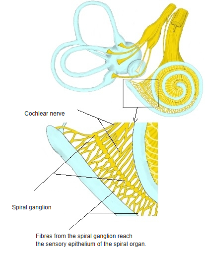
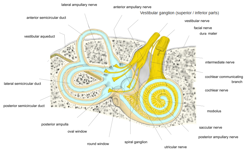
  • Its cell bodies form the spiral ganglion in the modiolus of the cochlea; their peripheral processes reach the spiral organ (organ of Corti).
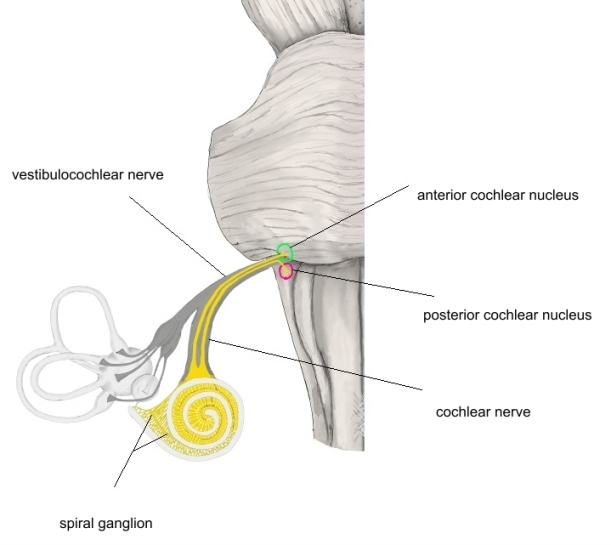
  • The central fibres enter the brainstem with the vestibular nerve and end in the ventral and dorsal cochlear nuclei.
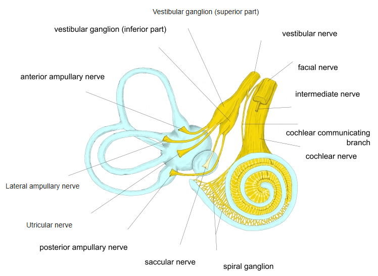
  • Inner ear showing the semicircular ducts, cochlea and the vestibular and cochlear nerves with their ganglia
    Inner ear and vestibulocochlear nerve
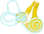
  • Membranous labyrinth showing the ampullary, utricular and saccular nerves, the vestibular ganglion and the cochlear nerve
    Branches of the vestibular and cochlear nerves
Course

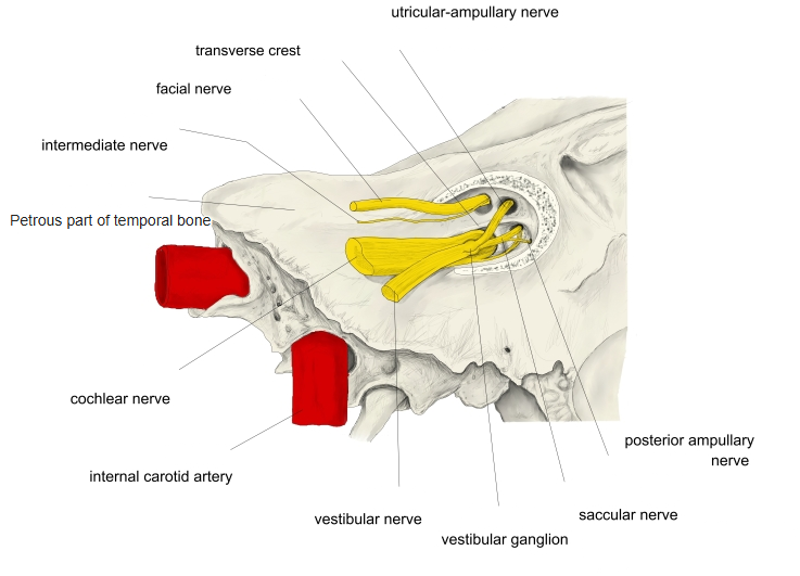
  • Passes through the internal acoustic meatus beside the vestibular and facial nerves.
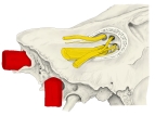
  • Petrous part of the temporal bone with the internal acoustic meatus and the facial, intermediate, cochlear and vestibular nerves
    Internal acoustic meatus
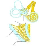
  • Inner ear with an enlarged view of the cochlea showing the spiral ganglion and the fibres of the cochlear nerve
    Cochlear nerve and spiral ganglion
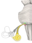
  • Brainstem and inner ear showing the cochlear nerve from the spiral ganglion to the anterior and posterior cochlear nuclei
    Cochlear nerve and cochlear nuclei
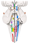
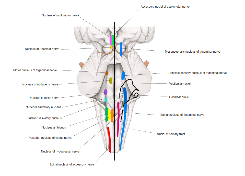
  • Posterior view of the brainstem with the motor nuclei on the left and the sensory nuclei on the right, each in its own colour
    Cranial nerve nuclei in the brainstem
References and sources

  1. Further reading: Cochlear nerve, Wikipedia.