Latin: nervus facialis
Overview
- The seventh cranial nerve (CN VII).
- Mixed nerve: motor fibres to the muscles of facial expression, plus the intermediate nerve with taste (sensory) and parasympathetic (secretory) fibres.
- A branchial (pharyngeal arch) nerve.
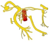
Facial nerve: nuclei and branches
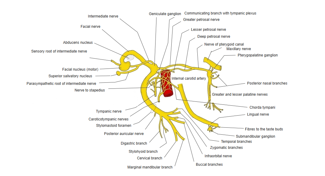
Origin
- Motor fibres: from the facial nucleus in the pons; they loop around the abducens nucleus (internal genu).
- Taste fibres: from cells of the geniculate ganglion, ending in the nucleus of the solitary tract.
- Parasympathetic fibres: from the superior salivatory nucleus.
- Leaves the brainstem between the pons and the medulla, medial to the vestibulocochlear nerve.
Course
- Enters the internal acoustic meatus with the vestibulocochlear nerve.
- Runs in the facial canal of the temporal bone; bends sharply backwards at the geniculum (geniculate ganglion), then descends.
- Leaves the skull through the stylomastoid foramen.
- Forms the parotid plexus inside the parotid gland (it does not supply the gland) and fans out to the face.
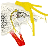
Facial nerve in the temporal bone 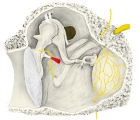
Walls of the tympanic cavity
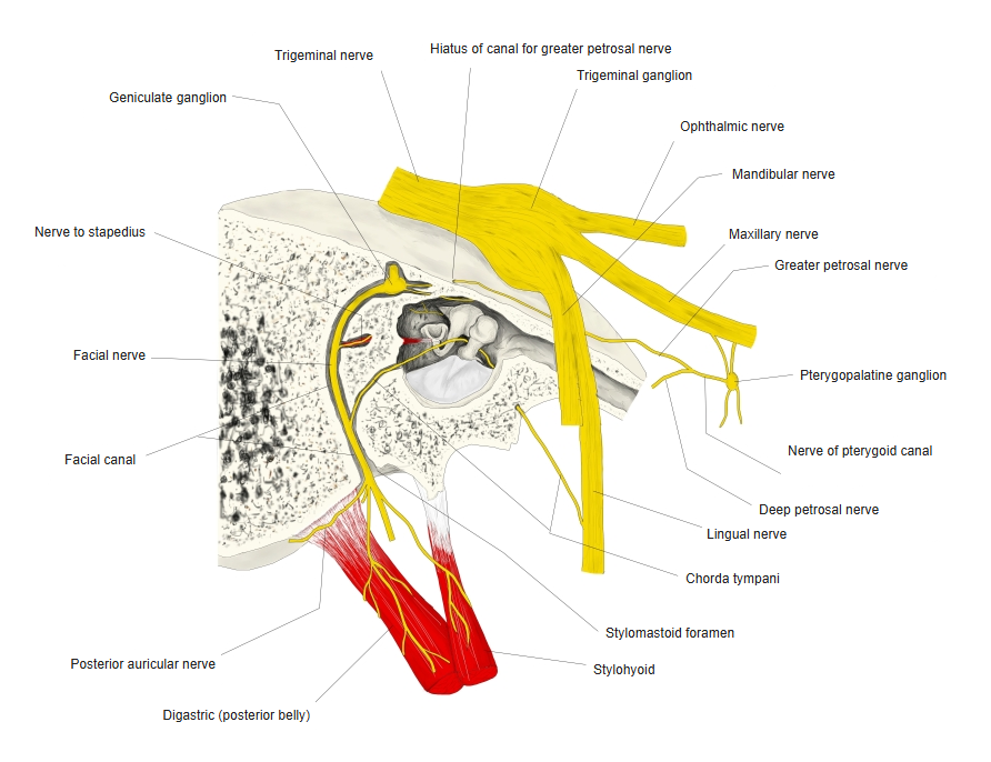
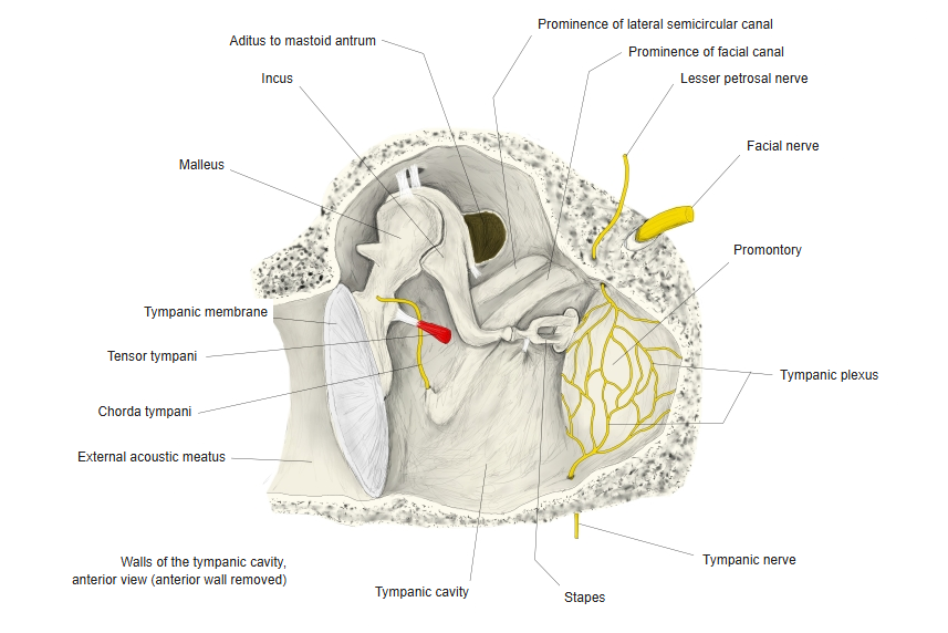
Branches and supply
| Branch | Supplies |
|---|---|
| In the facial canal | |
| Greater petrosal nerve | Parasympathetic fibres to the lacrimal gland and nasal glands (via the pterygopalatine ganglion) |
| Nerve to stapedius | Stapedius |
| Chorda tympani | Taste from the front two-thirds of the tongue; parasympathetic fibres to the submandibular and sublingual glands (joins the lingual nerve) |
| Communicating branch with the tympanic plexus | Tympanic plexus |
| After the stylomastoid foramen | |
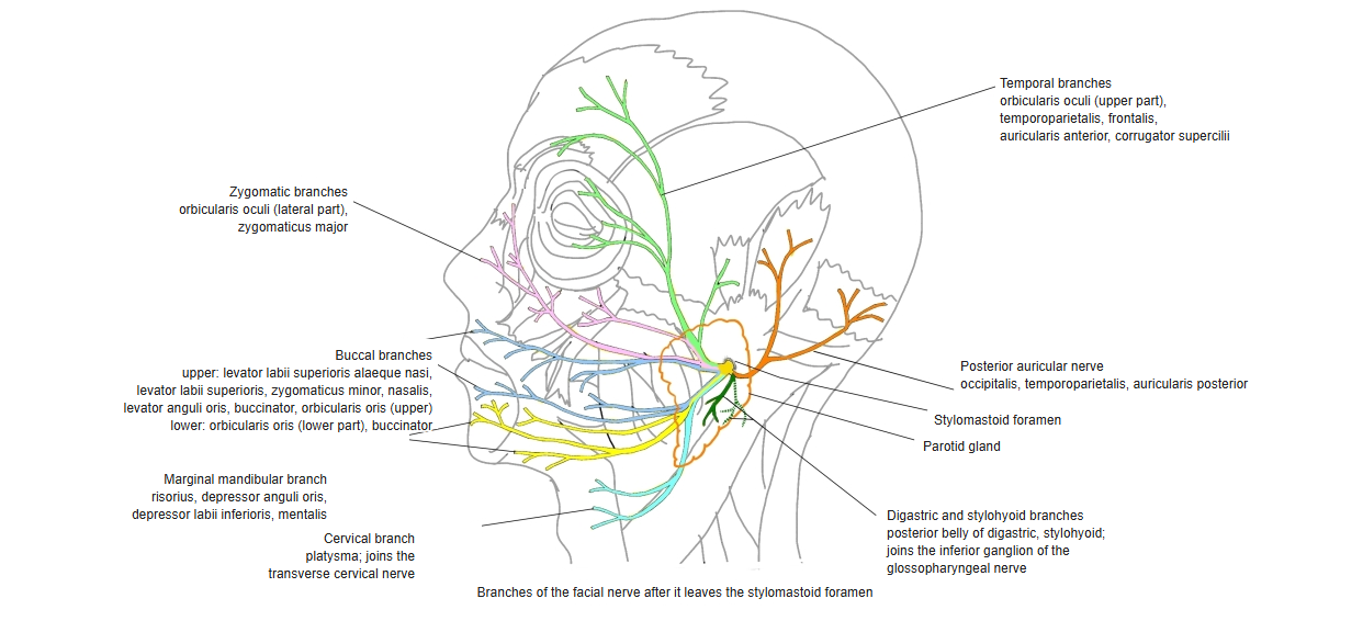
| Posterior auricular nerve | Occipitalis, auricularis posterior |
| Digastric branch and stylohyoid branch | Posterior belly of digastric, stylohyoid |
| From the parotid plexus | |
| Temporal branches | Frontalis, orbicularis oculi (upper part), corrugator supercilii, auricularis anterior |
| Zygomatic branches | Orbicularis oculi (lateral part), zygomaticus major |
| Buccal branches | Buccinator, orbicularis oris, zygomaticus minor, levator labii superioris, levator anguli oris, nasalis |
| Marginal mandibular branch | Risorius, depressor anguli oris, depressor labii inferioris, mentalis |
| Cervical branch | Platysma |
Branches to the muscles of the face
References and sources
- Further reading: Facial nerve, Wikipedia.