Latin: nervus vagus
Overview
- The tenth cranial nerve (X), the longest cranial nerve; a branchial (pharyngeal arch) nerve with motor, sensory and parasympathetic fibres.
- Its rootlets leave the medulla between the olive and the inferior cerebellar peduncle, just below the glossopharyngeal nerve.
- It supplies the pharynx and larynx and carries most of the parasympathetic supply of the thoracic and abdominal organs.
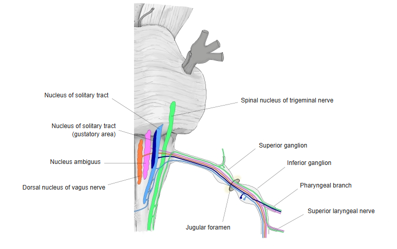
Nuclei and ganglia of the vagus nerve 
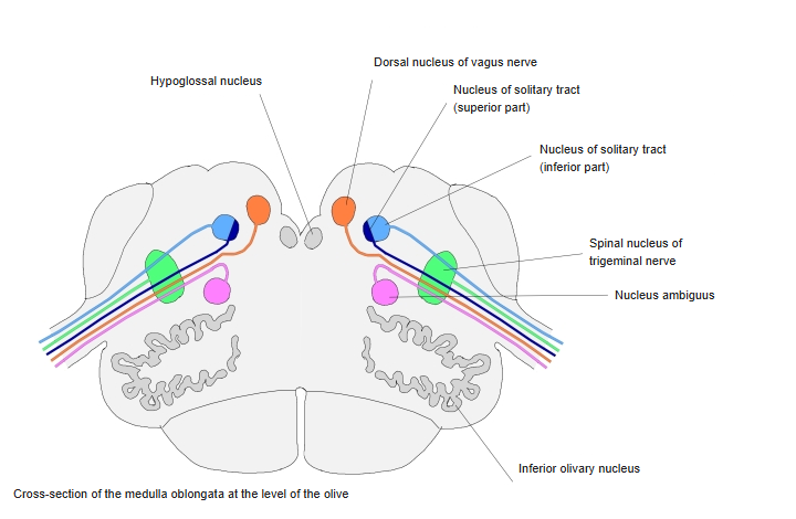
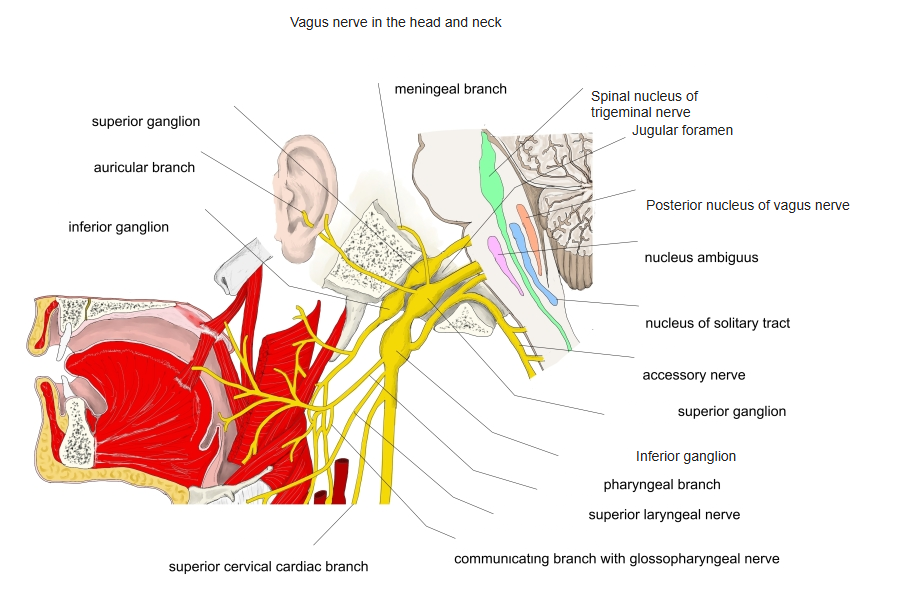
Vagal nuclei in the medulla oblongata
Nuclei and fibres
| Fibres | Nucleus |
|---|---|
| Parasympathetic | Dorsal nucleus of the vagus nerve: smooth muscle and glands of the viscera |
| Motor (branchial) | Nucleus ambiguus: skeletal muscles of the pharynx and larynx |
| Sensory | Cell bodies in the superior and inferior ganglia; central fibres to the spinal trigeminal nucleus (skin of the ear) and the nucleus of the solitary tract (viscera, taste) |
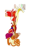
Course of the vagus nerve
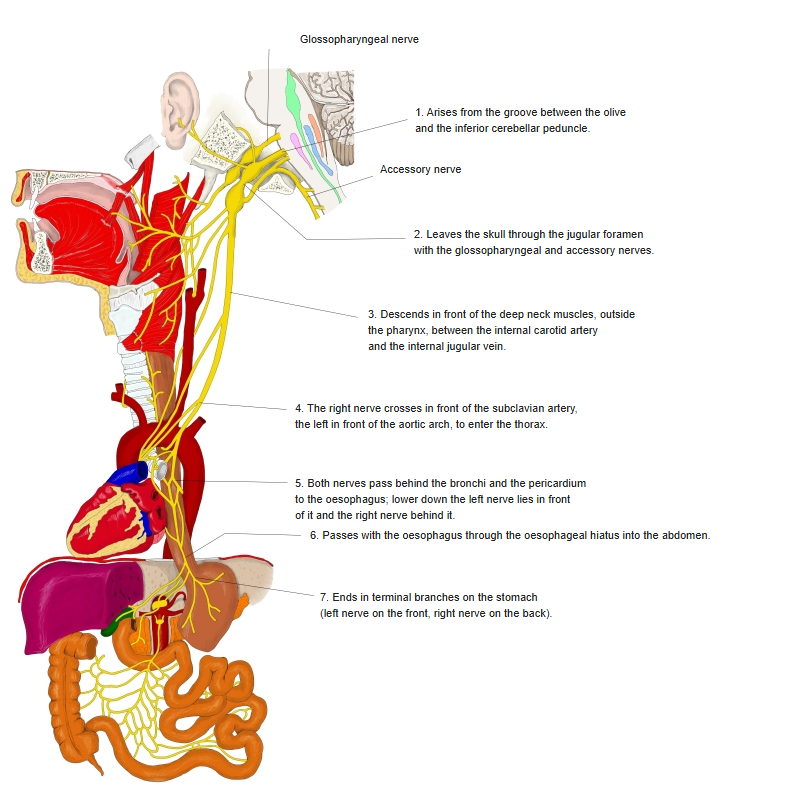
Course
- Leaves the skull through the jugular foramen with the glossopharyngeal and accessory nerves.
- Descends in the carotid sheath between the internal jugular vein and the internal and common carotid arteries.
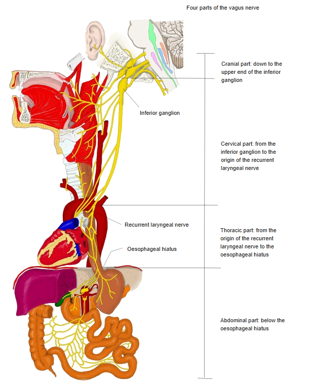
- The right vagus crosses in front of the subclavian artery, the left in front of the arch of the aorta, into the thorax.
- Both pass behind the root of the lung to the oesophagus; the left comes to lie in front of it, the right behind it.
- They pass through the oesophageal hiatus as the anterior and posterior vagal trunks and end on the stomach; branches reach the small and large intestine.
Ganglia
| Ganglion | Position | Connections |
|---|---|---|
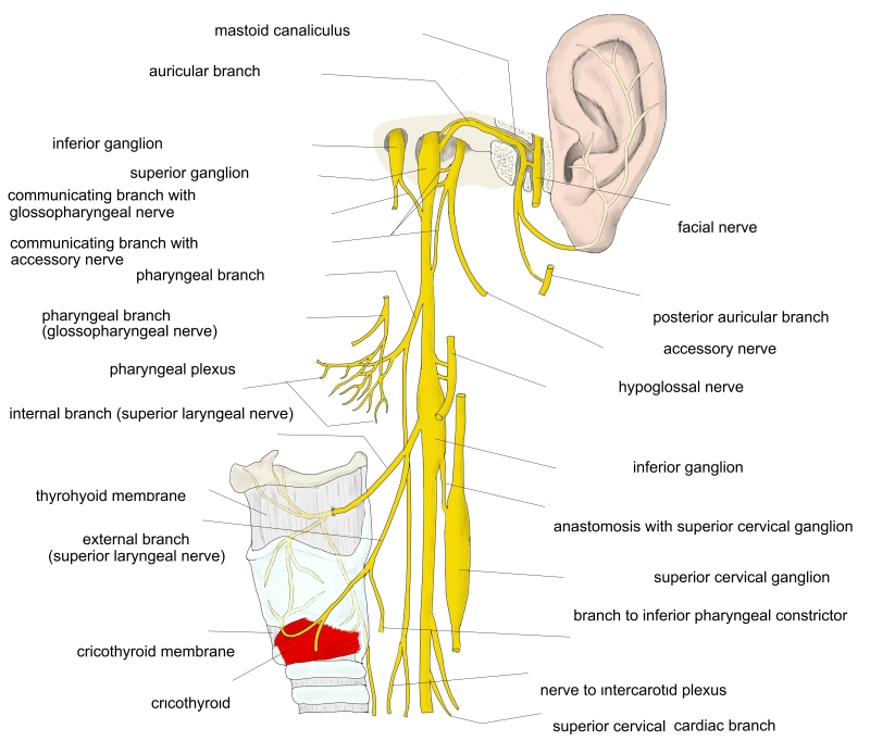
| Superior ganglion (jugular ganglion) | In the jugular foramen | Meningeal and auricular branches; communication with the superior cervical ganglion |
| Inferior ganglion (nodose ganglion) | Below the jugular foramen | Superior laryngeal nerve; communications with the superior cervical ganglion and the accessory nerve |
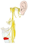
Branches of the vagus nerve in the neck 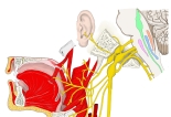
Vagus nerve in the head and neck
Branches
| Part | Branches |
|---|---|
| Head | Meningeal branch, auricular branch |
| Neck | Pharyngeal branches, superior laryngeal nerve, superior cervical cardiac branches, recurrent laryngeal nerve |
| Thorax | Thoracic cardiac branches, bronchial branches (pulmonary plexus), oesophageal branches (oesophageal plexus) |
| Abdomen | Gastric, hepatic, celiac branches, with renal and splenic branches |
Four parts of the vagus nerve
References and sources
- Further reading: Vagus nerve, Wikipedia.