Latin: musculus extensor digitorum
Overview
- One of the 11 muscles of the extensor group of the forearm, in the superficial layer.
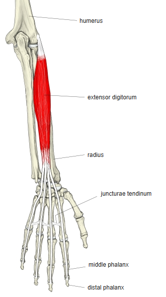
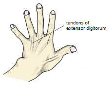
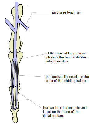
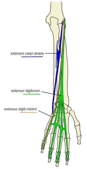
- Divides into four tendons which pass with the tendon of the extensor indicis through the 4th compartment of the extensor retinaculum; on the back of the hand they are linked by juncturae tendinum.
- Each tendon spreads into the dorsal digital expansion, reinforced by the interossei and lumbricals, and opposite the proximal interphalangeal joint divides into three slips: the central one to the middle phalanx, the two lateral ones uniting on the distal phalanx.
- Palpation: the tendons are easy to see on the back of the hand; two bellies can be felt under the skin.
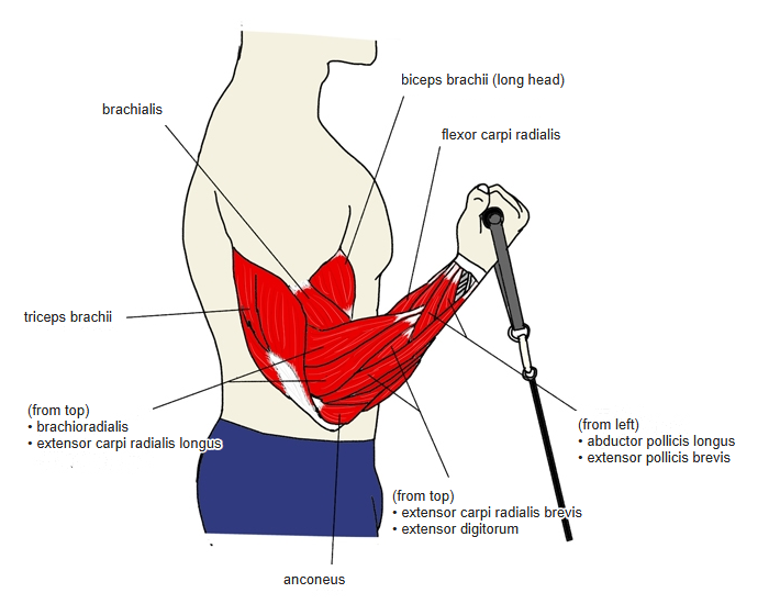
- Continuous in places with the extensor carpi ulnaris, extensor indicis, supinator, extensor carpi radialis brevis and extensor digiti minimi.
Position in the classification
Upper Limb › Forearm › Extensor group › Superficial posterior group
- Extensor digitorum
- Extensor digiti minimi
- Extensor carpi ulnaris
Illustrations
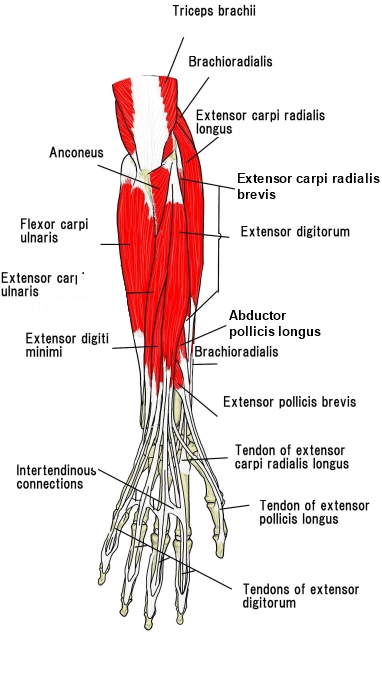
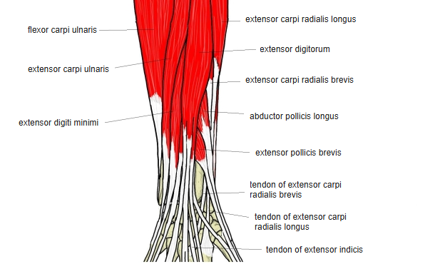
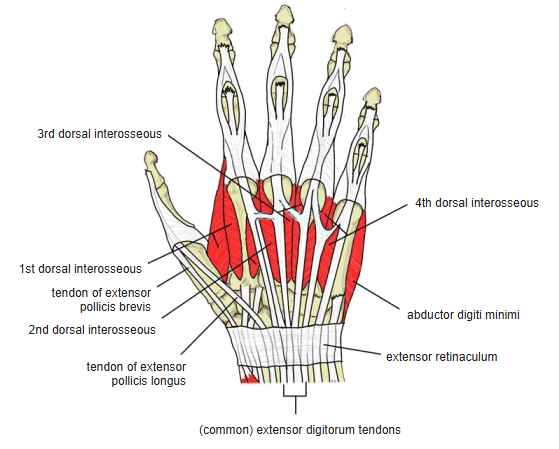
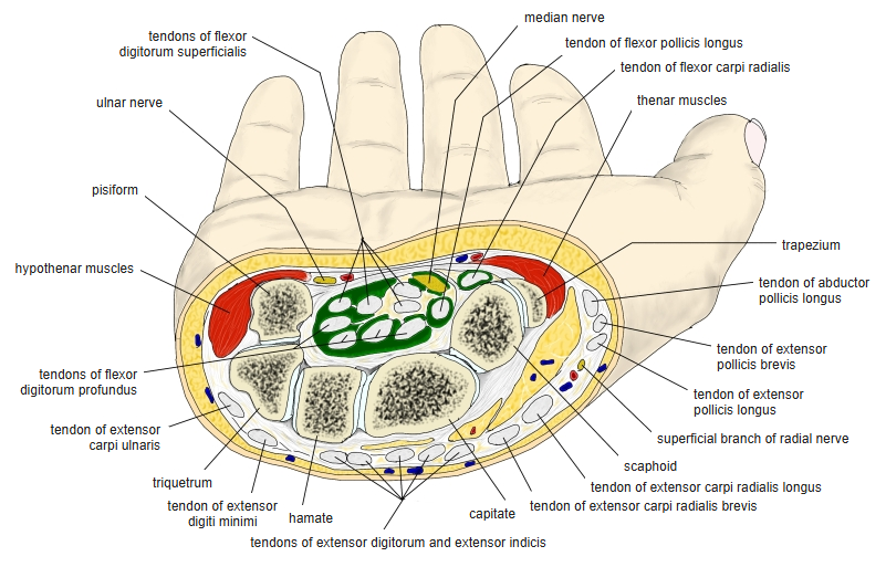
Origin / insertion
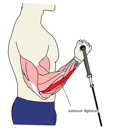
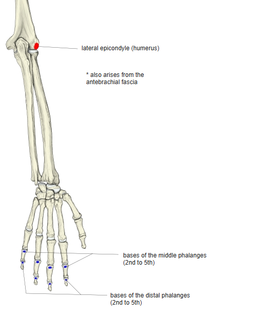
- Origin: lateral epicondyle of the humerus (common extensor tendon) and the antebrachial fascia
- Insertion: bases of the middle and distal phalanges of the 2nd to 5th fingers (via the dorsal digital expansion)
Action
- Extends the 2nd to 5th fingers
- Also extends the wrist
Innervation
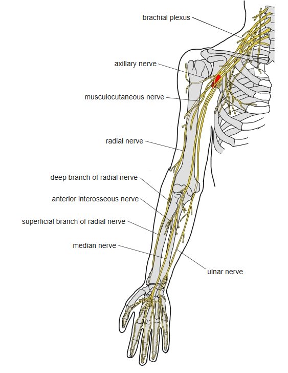
- Radial nerve, deep branch (posterior interosseous nerve), C7–C8; some sources C6–C8
Blood supply
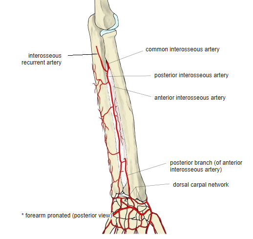
- Posterior interosseous artery
Anatomical variations
- An accessory tendon to the thumb may be present
- The tendon to the little finger (4th tendon) was missing in about 8% of male and 16% of female limbs in one study
Related terms
- Extensor indicis
- Extensor digiti minimi
- Extensor carpi ulnaris
- Lateral epicondyle
- Juncturae tendinum
- Dorsal digital expansion
- Extensor retinaculum
- Radial nerve
- Posterior interosseous artery
References and sources
- Further reading: Extensor digitorum muscle, Wikipedia.