Latin: musculus flexor digitorum profundus
Overview
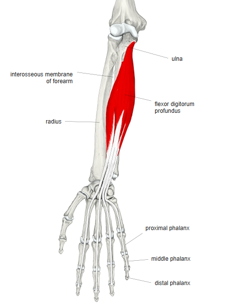
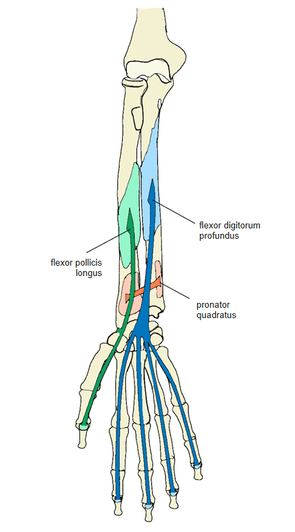
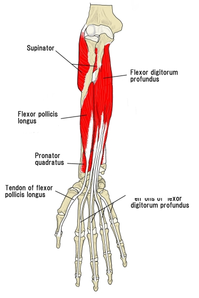
- One of the 8 flexor muscles of the forearm, in the deep layer with the flexor pollicis longus and pronator quadratus.
- Lies deep to the flexor digitorum superficialis, on the ulnar side of the forearm, over the ulnar half of the interosseous membrane.
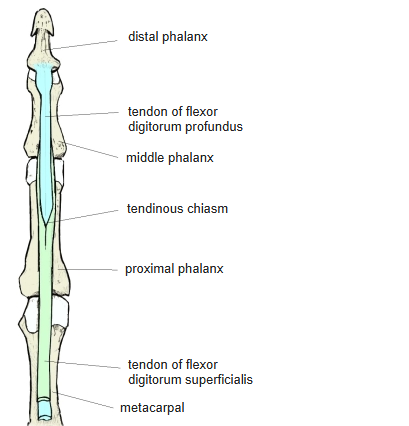
- Divides into four tendons that pass through the carpal tunnel and then through the split tendons of the flexor digitorum superficialis (hence also called the perforating muscle) to the distal phalanges.
- The lumbricals arise from its tendons: the 1st and 2nd from the radial sides of the tendons to the index and middle fingers, the 3rd and 4th from the adjacent sides of two tendons.
- Continuous in places with the biceps brachii, flexor carpi ulnaris, extensor and abductor pollicis longus, and flexor pollicis longus.
Position in the classification
Upper Limb › Forearm › Flexor group › Second and third layers
- Flexor digitorum superficialis
- Flexor digitorum profundus
- Flexor pollicis longus
Illustrations
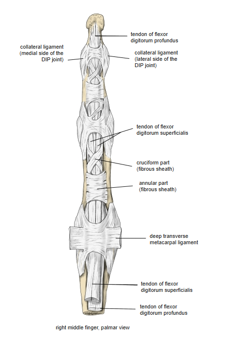
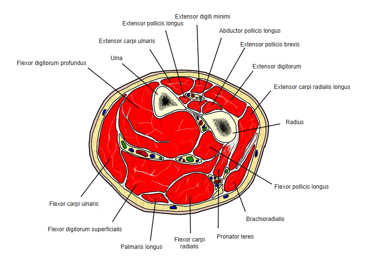
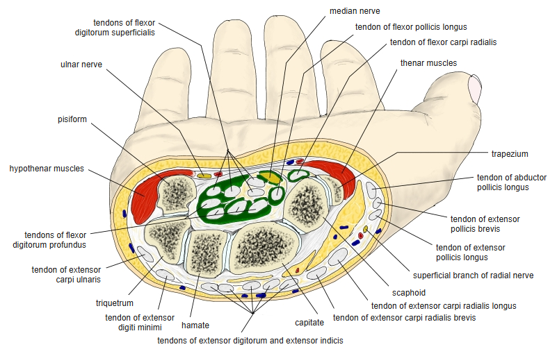
Origin / insertion
- Origin: upper three-quarters of the anterior and medial surfaces of the ulna, the medial side of the coronoid process and the ulnar half of the interosseous membrane (some sources: upper two-thirds of the anterior surface of the ulna)
- Insertion: bases of the distal phalanges of the 2nd to 5th fingers (palmar side)
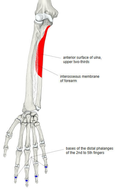
Action
- Flexes the distal interphalangeal (DIP) joints of the 2nd to 5th fingers; it also flexes the proximal interphalangeal and metacarpophalangeal joints of the same fingers
- Helps to flex the wrist
Innervation
- Lateral part (index and middle fingers): median nerve via the anterior interosseous nerve
- Medial part (ring and little fingers): ulnar nerve
- Spinal segments: C8, T1; some sources C7–T1
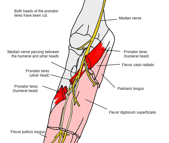
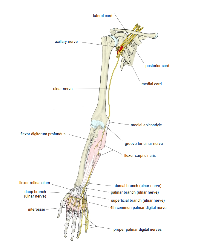
Blood supply
- Ulnar artery and anterior interosseous artery
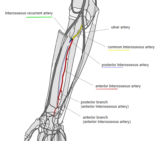
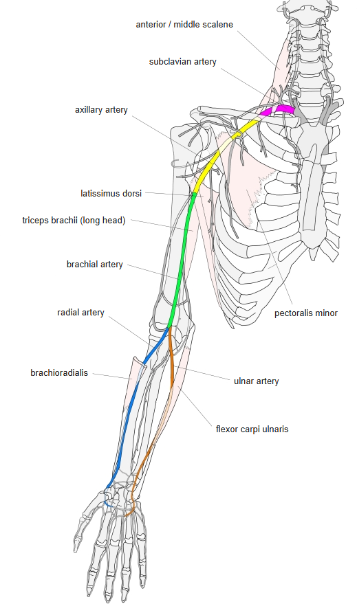
Anatomical variations
- The four bellies may be separate to varying degrees
- May also arise from the radius
- If the flexor digitorum superficialis lacks a tendon to a finger (especially the little finger), this muscle may provide an extra tendon, and vice versa
Related terms
- Flexor digitorum superficialis
- Flexor pollicis longus
- Pronator quadratus
- Lumbricals (hand)
- Interosseous membrane of forearm
- Carpal tunnel
- Median nerve
- Ulnar nerve
- Anterior interosseous artery
References and sources
- Further reading: Flexor digitorum profundus muscle, Wikipedia.