Latin: musculus rectus capitis posterior minor
Overview
- One of the 4 suboccipital muscles, in the second layer of the deep muscles of the back.
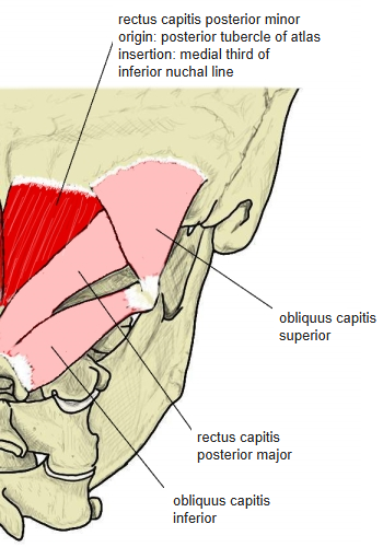
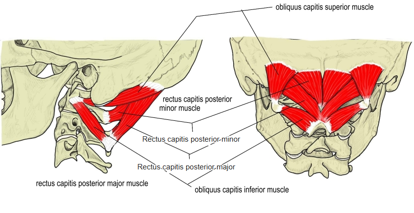
- A short muscle joining the atlas and the occipital bone, medial to the rectus capitis posterior major, which covers its lateral part.
- Starts as a narrow pointed tendon and widens as it ascends.
- Connective tissue links it to the spinal dura mater at the atlanto-occipital joint; these links have been proposed as one cause of cervicogenic headache.
Position in the classification
Trunk › Back › Deep back muscles › Second layer › Suboccipital muscles
- Rectus capitis posterior major
- Rectus capitis posterior minor
- Obliquus capitis inferior
- Obliquus capitis superior
Illustrations
Origin / insertion
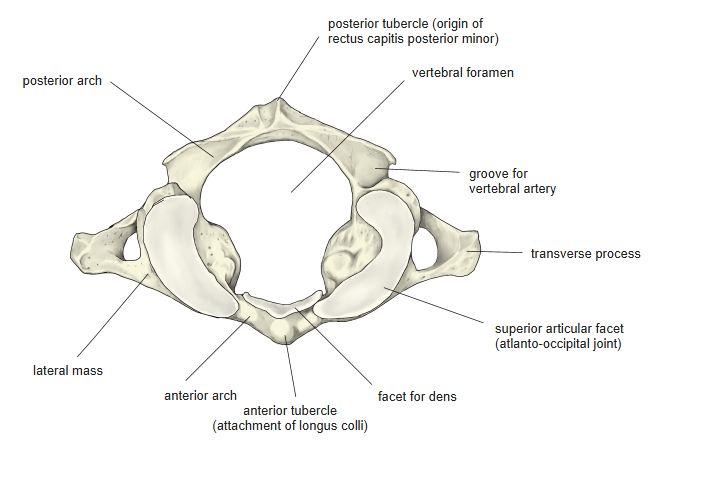
- Origin: posterior tubercle of the atlas
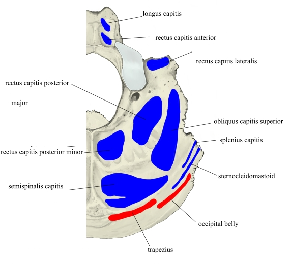
- Insertion: medial third of the inferior nuchal line of the occipital bone; English sources add the bone between this line and the foramen magnum, and the spinal dura mater
Action
- Draws the head backward (extension)
- Acting on one side, turns the head toward the same side
Innervation
- Suboccipital nerve (dorsal ramus of the 1st cervical nerve, C1)
Blood supply
- Not stated in the sources used here.
Anatomical variations
- An intermediate rectus capitis posterior may appear between the major and minor muscles
Related terms
- Suboccipital muscles
- Rectus capitis posterior major
- Obliquus capitis superior
- Obliquus capitis inferior
- Atlas
- Inferior nuchal line
- Dura mater
- Suboccipital nerve
References and sources
- Further reading: Rectus capitis posterior minor muscle, Wikipedia.