Latin: vesica urinaria
Overview
- Hollow muscular organ that stores urine; usual capacity about 300–500 ml.
- Lies in the front of the pelvis behind the pubic symphysis; when full it rises into the abdomen.
- Receives the two ureters and drains through the urethra.
- Its upper surface is covered by peritoneum; the rest lies outside the peritoneal cavity.
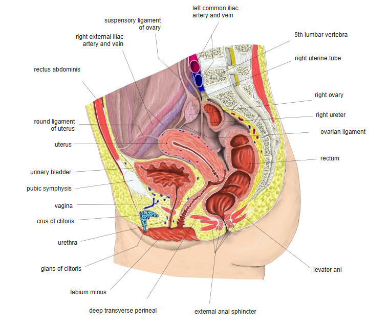
Female pelvis (median section)
Parts
| Part | Description |
|---|---|
| Apex | Points toward the pubic symphysis; joined to the umbilicus by the median umbilical ligament (urachus) |
| Body | Main part between apex and fundus |
| Fundus (base) | Posterior surface, receiving the ureters |
| Neck | Lowest part, continuous with the urethra |
| Trigone | Smooth triangle inside the base between the two ureteric orifices and the internal urethral orifice |
Relations and wall
- Male: behind lie the seminal vesicles, ductus deferens and rectum (rectovesical pouch); below is the prostate.
- Female: behind lie the uterus and vagina (vesicouterine pouch); the neck rests on the pelvic floor.
- Wall: transitional epithelium (urothelium) in folds that flatten as it fills; the smooth muscle is the detrusor.
- Arteries: superior and inferior vesical arteries (from the internal iliac artery; vaginal artery in the female).
- Nerves: parasympathetic pelvic splanchnic nerves (S2–S4) contract the detrusor for emptying; sympathetic fibres relax it.
References and sources
- Further reading: Urinary bladder, Wikipedia.