Latin: ureter
Overview
- Paired muscular tube carrying urine from the renal pelvis to the urinary bladder.
- About 25–30 cm long and 3–4 mm in diameter.
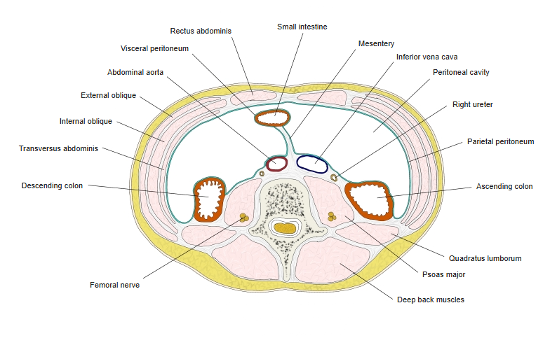
- Retroperitoneal along its whole course.
- Described in three parts: abdominal, pelvic and intramural (within the bladder wall).
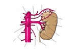
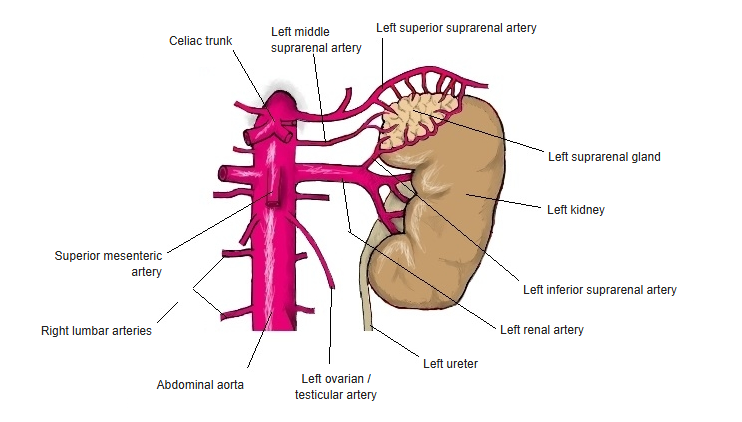
Left kidney, suprarenal gland and ureter
Course and relations
- Leaves the kidney at the hilum and descends on the front of psoas major.
- Crossed in front by the testicular (ovarian) vessels.
- Enters the pelvis by crossing the bifurcation of the common iliac artery (or the start of the external iliac artery).
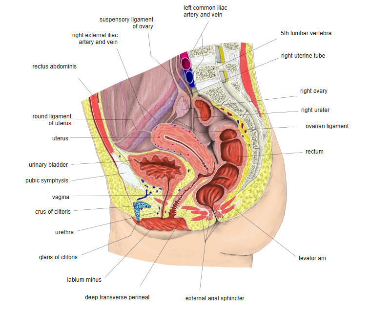
- Runs down the lateral pelvic wall, then turns forward and medially to the bladder.
- In the male the ductus deferens crosses above it; in the female the uterine artery crosses above it near the cervix.
- Passes obliquely through the bladder wall and opens at the ureteric orifice.
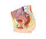
Female pelvis (median section) 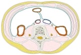
Transverse section at the level of L4
Physiological narrowings
| Site | Note |
|---|---|
| 1. Pelviureteric junction | Where the renal pelvis becomes the ureter |
| 2. Pelvic brim | Where the ureter crosses the common iliac vessels |
| 3. Intramural part | Where the ureter passes through the bladder wall – the narrowest point |
- Urinary stones most often lodge at these three points and cause colicky pain radiating from the loin to the groin.
Wall and function
- Mucosa: transitional epithelium (urothelium) in longitudinal folds, giving a star-shaped lumen.
- Muscular layer: smooth muscle, inner longitudinal and outer circular, with an extra outer longitudinal layer in the lower third.
- Adventitia: fibrous outer coat carrying vessels and nerves.
- Peristalsis carries urine to the bladder in small spurts.
- Because the ureter runs obliquely through the bladder wall, a filling bladder compresses it and prevents reflux of urine.
Blood supply and nerves
- Arteries: small branches from the renal artery, testicular or ovarian artery, abdominal aorta, common iliac, internal iliac, vesical and (female) uterine arteries.
- Nerves: autonomic fibres from the renal, aortic and hypogastric plexuses; pain fibres reach the spinal cord at about T11–L2.
References and sources
- Further reading: Ureter, Wikipedia.