Latin: ductus cochlearis
Overview
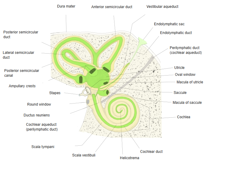
- The part of the membranous labyrinth inside the cochlea; also called the scala media.
- A blind-ended spiral tube, triangular in cross-section, filled with endolymph.
- Joined to the saccule by the ductus reuniens; its apical end is closed (cecum cupulare).
- Contains the organ of hearing, the spiral organ (organ of Corti).
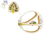
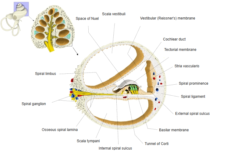
Cochlea, cross-section of one turn 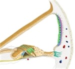
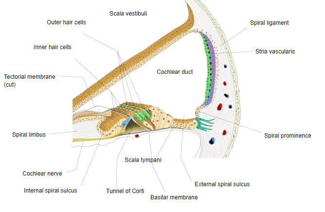
Cochlear duct, cross-section
Walls
| Wall | Structure | Notes |
|---|---|---|
| Roof (vestibular wall) | Vestibular (Reissner) membrane | Very thin; separates the duct from the scala vestibuli |
| Outer wall | Stria vascularis on the spiral ligament | Vascular epithelium that produces endolymph; the spiral prominence lies below it |
| Floor (tympanic wall) | Basilar membrane and the osseous spiral lamina | Carries the spiral organ; separates the duct from the scala tympani |
- The spiral limbus on the osseous spiral lamina gives attachment to the tectorial membrane, which lies over the hair cells.
- The internal spiral sulcus lies between the limbus and the spiral organ, the external spiral sulcus lateral to it.
Labyrinth in the temporal bone (schematic)
References and sources
- Further reading: Cochlear duct, Wikipedia.