Latin: iris
Overview
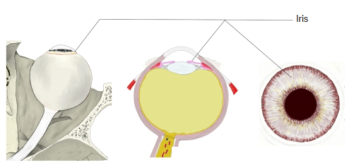
- The front part of the vascular layer: a thin, pigmented, contractile disc in front of the lens.
- Its central opening is the pupil; by changing its size the iris controls how much light enters the eye.
- Divides the space between the cornea and lens into the anterior and posterior chambers; its root (ciliary margin) is attached to the ciliary body.
- Its colour depends on the amount of melanin in the stroma: little pigment gives blue or grey eyes, much gives brown; the pigment epithelium at the back is always dark.
Iris in three views 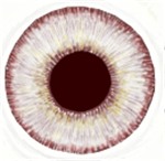
Iris and pupil
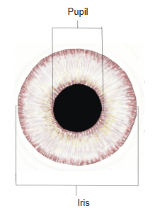
Anterior surface
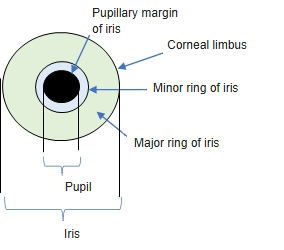
- The pupillary margin borders the pupil; the ciliary margin joins the ciliary body.
- A wavy ridge, the collarette, divides the surface into an inner pupillary zone (minor ring) and an outer ciliary zone (major ring).
- Between the iris and the cornea, at the periphery, lies the iridocorneal angle, where the aqueous humor drains.
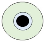
Iris, anterior view (diagram) 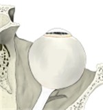
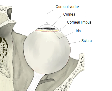
Front of the eyeball
Layers
| Layer (front to back) | Features |
|---|---|
| Anterior border layer | Condensed stroma with fibroblasts and pigment cells; no true epithelium |
| Stroma | Loose, vascular connective tissue with pigment cells; contains the sphincter pupillae near the pupillary margin |
| Dilator pupillae | Radial myoepithelial cells forming the anterior layer of the epithelium |
| Pigment epithelium | Densely pigmented posterior layer (iridial part of the retina); stops light passing through the iris |
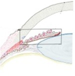
Iris, section 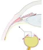
Iris and ciliary body, section
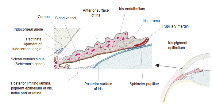
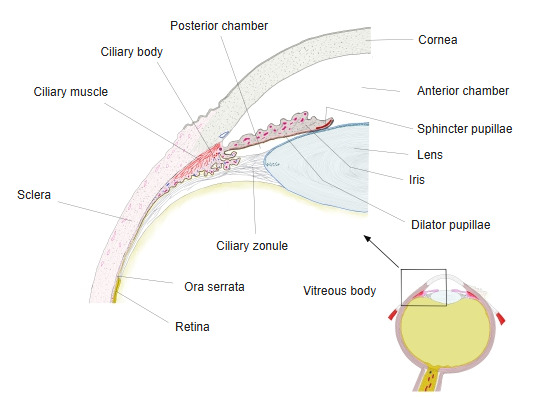
Vessels and nerves
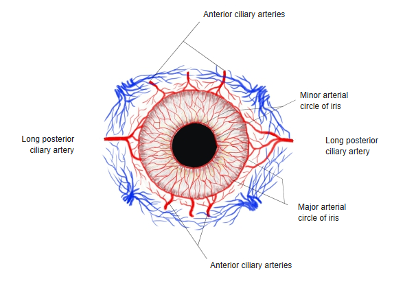
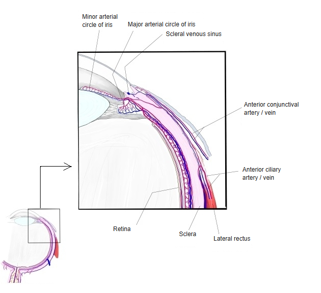
- Major arterial circle: at the ciliary margin, formed by the long posterior ciliary and anterior ciliary arteries.
- Minor arterial circle: near the collarette, joined to the major circle by radial vessels.
- Veins run with the arteries and drain mainly into the vorticose veins.
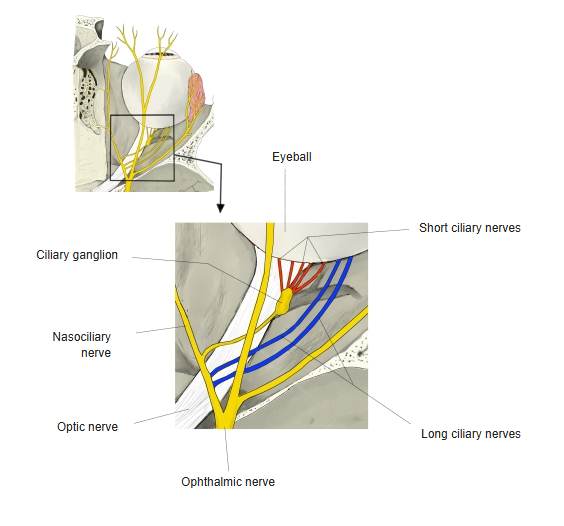
- Parasympathetic fibres (short ciliary nerves, from the ciliary ganglion) supply the sphincter pupillae.
- Sympathetic fibres (mainly via the long ciliary nerves) supply the dilator pupillae.
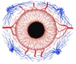
Arteries of the iris 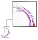
Arterial circles of the iris, section 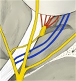
Ciliary nerves
Muscles of the iris
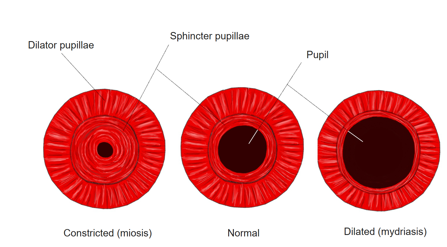
- Sphincter pupillae: a ring of smooth muscle around the pupil; it narrows the pupil (miosis).
- Dilator pupillae: radial fibres; it widens the pupil (mydriasis).
Size of the pupil
References and sources
- Further reading: Iris, Wikipedia.