Latin: sacculus
Overview
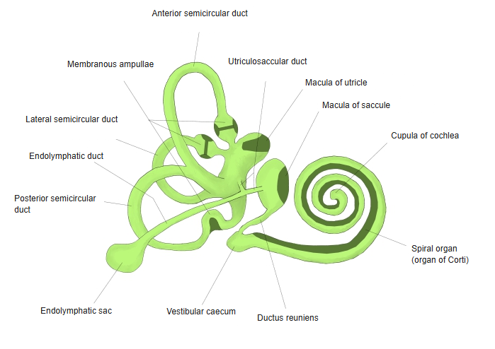
- The smaller, rounded membranous sac in the vestibule, lying in the spherical recess in front of the utricle.
- Its sensory area, the macula of the saccule, lies roughly vertical on its medial wall and senses vertical linear acceleration and head position.
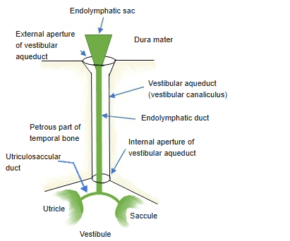
- Connected below to the cochlear duct by the ductus reuniens and to the utricle by the utriculosaccular duct.
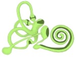
Membranous labyrinth 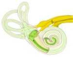
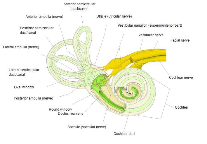
Labyrinth and its nerves
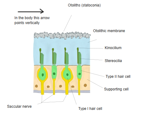
Structure of the macula
- Built like the macula of the utricle: hair cells and supporting cells covered by an otolithic membrane carrying otoliths.
- The two maculae lie roughly at right angles, so together they detect linear acceleration in all directions.
- Fibres from the macula run in the saccular nerve to the inferior part of the vestibular ganglion and on in the vestibular nerve.
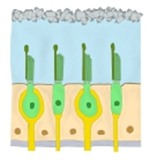
Macula, structure 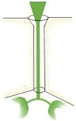
Endolymphatic duct and sac (schematic)
References and sources
- Further reading: Saccule, Wikipedia.