Latin: corpus vitreum
Overview
- A clear gel that fills the space between the lens and the retina (vitreous chamber).
- Makes up about four-fifths of the volume of the eyeball (about 4 ml); a sphere slightly flattened from front to back.
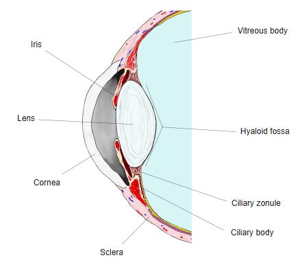
- In front it has a hollow for the lens, the hyaloid fossa.
- About 99% water with a fine network of collagen fibrils and hyaluronan; it transmits light and supports the retina.
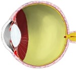
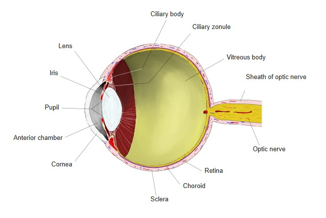
Eyeball, longitudinal section 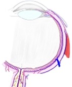
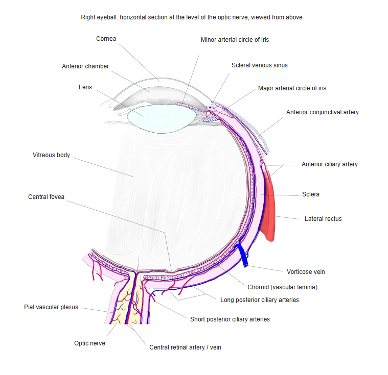
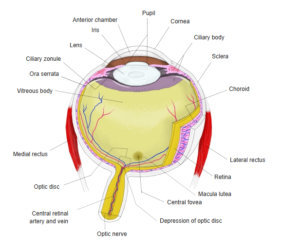
Right eyeball, horizontal section
Features
- Its surface is condensed into a thin vitreous membrane (hyaloid membrane).
- It is attached most firmly at the ora serrata (vitreous base) and around the optic disc.
- A narrow hyaloid canal runs from the optic disc to the back of the lens – the remnant of the fetal hyaloid artery.
- With age the gel partly liquefies; shrinking may cause floaters or detach it from the retina (posterior vitreous detachment).
Lens in place, section 
Eyeball, horizontal section
References and sources
- Further reading: Vitreous body, Wikipedia.