Latin: bulbus oculi
Overview
- The organ of sight: the central part of the visual organ, which receives light and turns it into nerve signals.
- Sits in the front part of the bony orbit, cushioned by orbital fat; below and laterally it projects slightly beyond the orbital margin.
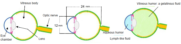
- Almost a sphere, about 24 mm across, with the transparent cornea bulging a little at the front.
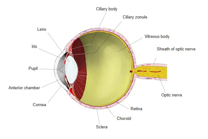
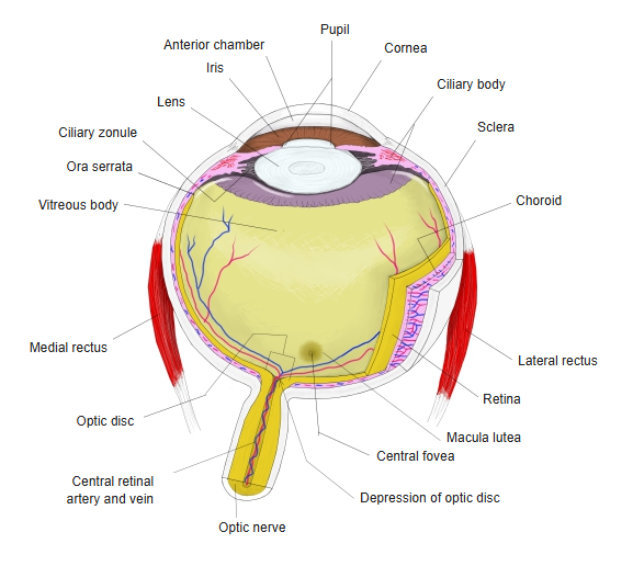
- Its wall has three layers – fibrous, vascular and inner (nervous) – around a transparent interior: aqueous humor, lens and vitreous body.
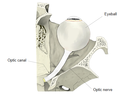
- Behind, it continues into the optic nerve; six extraocular muscles move it.
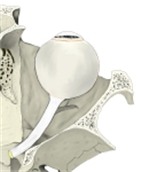
Eyeball and optic nerve in the orbit 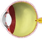
Eyeball, longitudinal section
Shape, size and axes
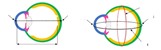
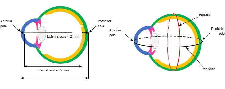
- Anterior pole: the centre of the corneal curvature; posterior pole: the opposite point at the back.
- External axis: the line from the anterior to the posterior pole on the outer surface – about 24 mm in the adult.
- Internal axis: the same line measured from the back of the cornea to the inner surface of the retina – about 22 mm.
- Equator: the largest circumference, midway between the poles; meridians run from pole to pole.
- Width (transverse diameter) and height (vertical diameter) are each close to 24 mm; individual differences are only 1–2 mm.
- At birth the eyeball measures about 16–17 mm; it reaches nearly adult size in childhood.
- The visual axis (line of sight) runs from the object seen to the central fovea and makes a small angle with the optic axis.
Poles, axes and equator 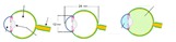
Size and contents (diagram)
Wall of the eyeball
| Layer | Parts | Main features |
|---|---|---|
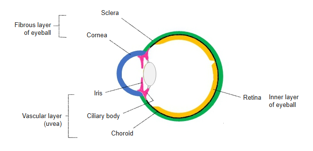
| Fibrous layer (outer) | Sclera and cornea | Tough white sclera over the posterior five-sixths; the transparent, more curved cornea in front. Keeps the shape of the eye and protects it |
| Vascular layer (middle; uvea) | Choroid, ciliary body, iris | Rich in blood vessels and pigment. The ciliary muscle changes the shape of the lens; the iris adjusts the size of the pupil |
| Inner layer (retina) | Optic part and blind (non-visual) part of the retina | The optic part contains the photoreceptors (rods and cones); it ends forward at the ora serrata. The blind part lines the ciliary body and iris |
- The sclera and cornea meet at the corneoscleral junction (limbus), where the scleral venous sinus drains the aqueous humor.
- The optic nerve leaves the eyeball through the lamina cribrosa of the sclera, slightly medial to the posterior pole.
- On the retina, the optic disc (where the nerve fibres leave; the blind spot) lies medial to the macula lutea, whose central pit, the central fovea, gives the sharpest vision.
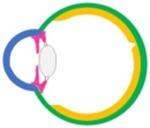
Three layers of the wall (diagram) 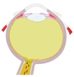
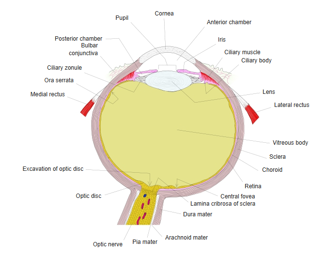
Right eyeball, horizontal section (model)
Contents of the eyeball
| Structure | Description |
|---|---|
| Anterior chamber | Space between the cornea and the iris, filled with aqueous humor |
| Posterior chamber | Narrow space between the iris and the lens; the two chambers communicate through the pupil |
| Aqueous humor | Clear watery fluid made by the ciliary processes; flows from the posterior to the anterior chamber and drains at the iridocorneal angle. Maintains the intraocular pressure (about 12–22 mmHg) |
| Lens | Transparent biconvex disc behind the iris, held by the ciliary zonule; its curvature changes for near and far vision (accommodation) |
| Vitreous body | Clear gel filling the space behind the lens – about four-fifths of the volume of the eyeball |
- The lens divides the interior into a smaller anterior segment (the chambers) and a larger posterior segment (the vitreous body).
- Light passes through the cornea, aqueous humor, pupil, lens and vitreous body before it reaches the retina; the cornea does most of the focusing.
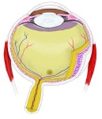
Eyeball, horizontal section
Movements and extraocular muscles
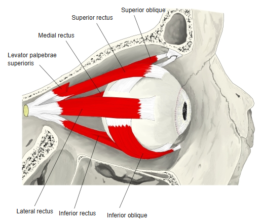
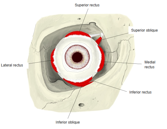
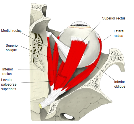
- Six extraocular muscles turn the eyeball: four recti – superior, inferior, medial and lateral rectus – and two obliques – superior and inferior oblique.
- The four recti arise from the common tendinous ring at the apex of the orbit and insert on the sclera in front of the equator.
- The obliques insert behind the equator; the superior oblique reaches the eyeball through the trochlea at the upper medial margin of the orbit.
- The levator palpebrae superioris lies above the superior rectus and raises the upper eyelid; it does not move the eyeball.
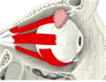
Right extraocular muscles, lateral view 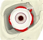
Right extraocular muscles, anterior view 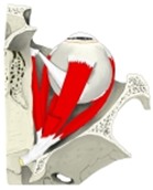
Right extraocular muscles, superior view
Blood supply, lymph and nerves
- Arteries: branches of the ophthalmic artery – the central retinal artery for the inner retina, and the short and long posterior ciliary and anterior ciliary arteries for the choroid, ciliary body and iris.
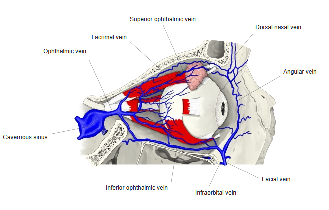
- Veins: the central retinal vein runs with its artery; the vorticose veins (usually 4–6) collect most of the blood of the vascular layer near the equator, pierce the sclera and drain into the superior and inferior ophthalmic veins.
- Lymph: the eyeball has no true lymphatic vessels; fluid moves through tissue spaces such as the chambers, the perichoroidal space and the space around the optic nerve.
- Optic nerve: carries the visual fibres from the retina to the brain.
- Long ciliary nerves: sensory branches of the nasociliary nerve (ophthalmic nerve, CN V1); they pierce the sclera near the optic nerve and supply the cornea, iris and ciliary body.
- Short ciliary nerves: from the ciliary ganglion; they carry sensory, sympathetic and parasympathetic fibres to the sphincter pupillae, dilator pupillae and ciliary muscle.
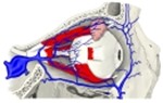
Veins of the orbit 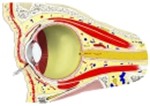
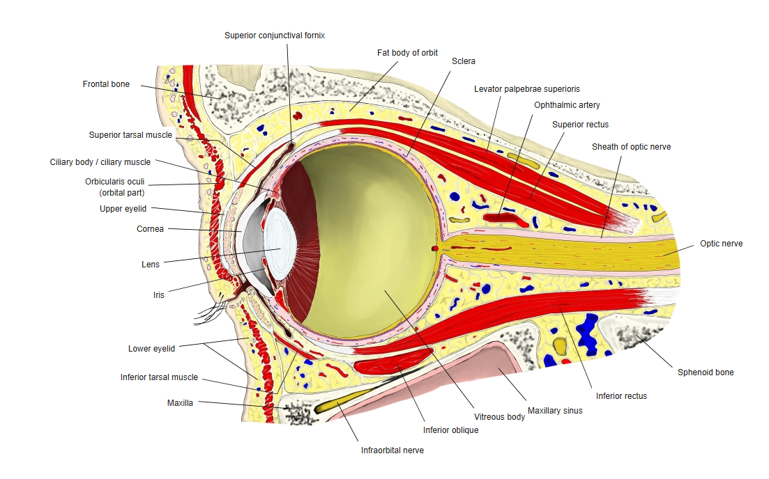
Orbit, longitudinal section
References and sources
- Further reading: Eyeball, Wikipedia.