Latin: Splen (Lien)
Overview
- The largest lymphoid organ; it filters the blood.
- Lies in the left upper abdomen under the 9th to 11th ribs, behind the stomach.
- About the size of a fist (roughly 12 × 7 × 3 cm, about 150 g in the adult).
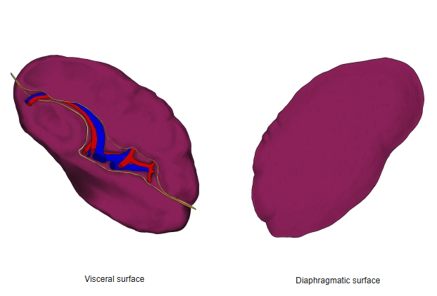
Surfaces and hilum
- Diaphragmatic surface: convex and smooth, facing the diaphragm.
- Visceral surface: concave, with impressions for the stomach, left kidney and left colic flexure.
- The hilum on the visceral surface transmits the splenic vessels; the spleen is covered by peritoneum and is held by the gastrosplenic and splenorenal ligaments.
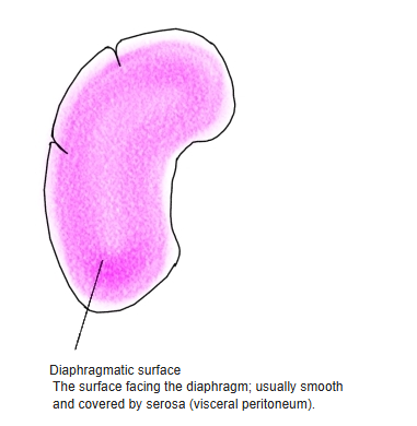
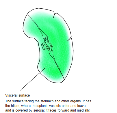
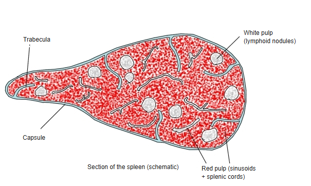
Internal structure
- A fibrous capsule sends trabeculae into the organ.
- White pulp: lymphoid tissue around the arteries (lymphoid nodules, periarterial sheaths); starts immune responses to blood-borne antigens.
- Red pulp: venous sinusoids and splenic cords; removes old red blood cells and stores platelets.
Blood vessels
- Artery: the splenic artery from the celiac trunk, dividing into several branches at the hilum (splenic segments).
- Vein: the splenic vein, which joins the superior mesenteric vein to form the portal vein.
Clinical notes
- The spleen is the abdominal organ most often injured in blunt trauma; rupture causes serious internal bleeding.
- After removal of the spleen (splenectomy), patients are more prone to infections by encapsulated bacteria.
- An enlarged spleen (splenomegaly) can be felt below the left costal margin.
References and sources
- Further reading: Spleen, Wikipedia.