Latin: Thymus
Overview
- A primary lymphoid organ where T lymphocytes mature.
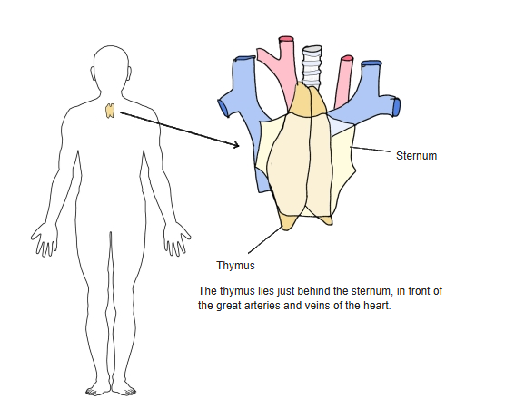
- Lies in the superior and anterior mediastinum, behind the sternum and in front of the great vessels of the heart.
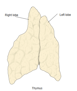
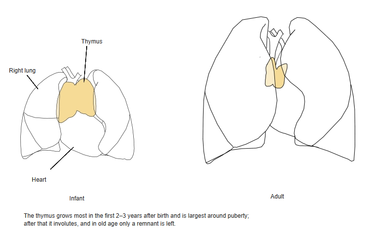
- Made of right and left lobes; it is largest relative to body size in early childhood and largest in absolute size around puberty, then is gradually replaced by fat.
Changes with age
- Grows rapidly in the first years of life.
- After puberty it involutes; in the elderly only a small remnant surrounded by fat is left.
Internal structure
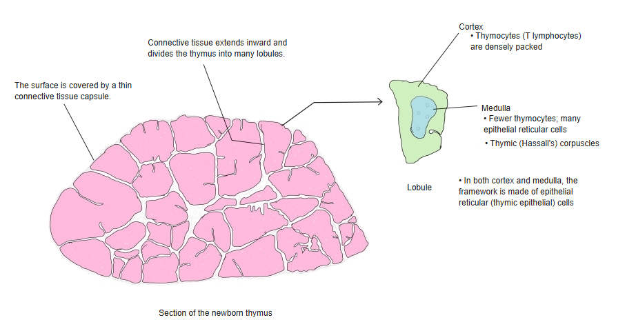
- A thin connective tissue capsule sends septa inward, dividing each lobe into lobules.
- Cortex: densely packed thymocytes (developing T cells).
- Medulla: fewer thymocytes, more epithelial reticular cells and the thymic (Hassall's) corpuscles.
Blood vessels
- Arteries: thymic branches of the internal thoracic artery and the inferior thyroid arteries.
- Veins: thymic veins to the left brachiocephalic vein and the internal thoracic veins.
Clinical notes
- Tumours of the thymus (thymomas) are often linked with myasthenia gravis.
- Absence of the thymus (as in DiGeorge syndrome) causes severe immune deficiency.
References and sources
- Further reading: Thymus, Wikipedia.