Latin: Venae pulmonales
Overview
- The veins that carry oxygen-rich blood from the lungs to the heart.
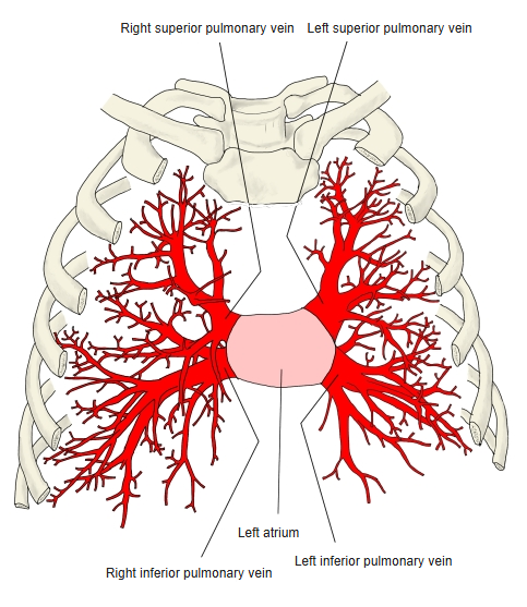
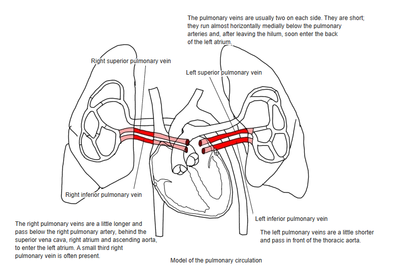
- Usually four in number: right superior, right inferior, left superior and left inferior.
- Open into the back of the left atrium.
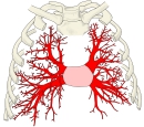
Pulmonary veins (front view) 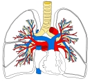
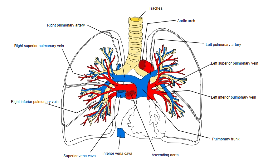
Pulmonary arteries and veins
Course
- Begin in the capillary networks of the alveoli; small veins run between the lung segments (intersegmental veins).
- At the hilum, the pulmonary veins lie in front of and below the pulmonary artery and bronchus.
- The right middle lobe vein usually joins the right superior pulmonary vein.
Clinical notes
- Muscle sleeves around the pulmonary vein openings can trigger atrial fibrillation; catheter ablation isolates them.
- The number of veins varies (three or five is not rare).
References and sources
- Further reading: Pulmonary veins, Wikipedia.